
Multi-eye stereo imaging microscope and imaging method thereof

A stereoscopic imaging and imaging method technology, applied in the field of biomedical microscopic imaging, can solve problems such as incomplete stereo matching algorithms, inaccurate stereoscopic imaging effects, and inability to capture images from multiple angles at the same time, achieving low cost and short consumption when the effect

Inactive Publication Date: 2020-01-24
SOUTHEAST UNIV
View PDF7 Cites 2 Cited by
  • Summary
  • Abstract
  • Description
  • Claims
  • Application Information

AI Technical Summary

Problems solved by technology

However, this method also has difficulties to be solved in many aspects: 1) the existing stereo microscope has only two eyepiece tubes, which cannot capture images from multiple angles at the same time; 2) under the size of the microscope, the multi-CCD electronic eyepiece The camera calibration algorithm is different from the multi-eye camera calibration in the macro scene, and a special algorithm needs to be proposed for it; 3) The specific stereo matching algorithm for microscopic samples is not yet perfect, and the stereo imaging effect is not accurate enough

Method used


Image

  • Multi-eye stereo imaging microscope and imaging method thereof
  • Multi-eye stereo imaging microscope and imaging method thereof
  • Multi-eye stereo imaging microscope and imaging method thereof

Examples


Embodiment Construction

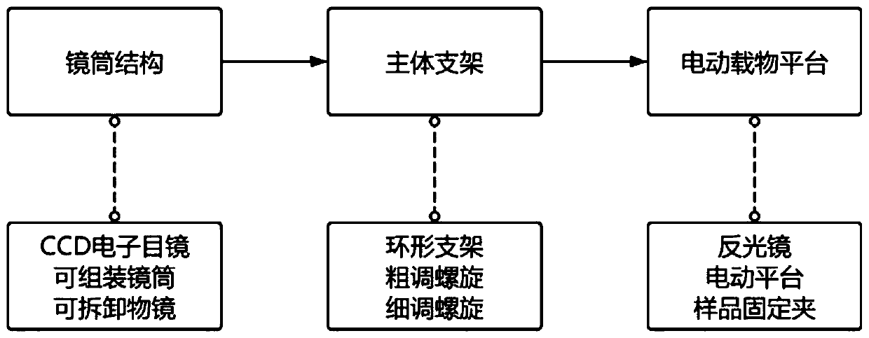
[0026] like figure 1 As shown, a multi-eye stereoscopic imaging microscope includes: a lens barrel structure, a microscope main body support structure and a motorized object platform; the microscope main body support structure is used to clamp and fix each lens barrel structure, and adjust the work between each lens barrel Distance; the right side of the annular bracket in the microscope main bracket structure is provided with a grip arm, and the grip arm is connected with the electric object loading platform through a hinge support.

[0027] The lens barrel structure includes a CCD electronic eyepiece, an assembling lens barrel and a detachable objective lens; the CCD electronic eyepiece is used to collect the optical signal transmitted by the assembling lens barrel, convert it into an electrical signal, and upload it to the PC host through the USB data cable machine; the assembleable lens barrel is used to connect the CCD electronic eyepiece and the detachable objective lens...


PUM


Abstract

The invention discloses a multi-eye stereo imaging microscope and an imaging method thereof. The multi-eye stereo imaging microscope includes a lens barrel structure, a microscope main body supportingstructure, and an electric objective table. The imaging method of the multi-eye stereo imaging microscope comprises the following steps of: (1) shooting a checkerboard calibration board, and using atwo-step calibration method to perform a camera calibration on multi-eye CCD electronic eyepieces to determine the relative position of each eyepiece; (2), performing image registration on a capturedsample according to the camera parameters obtained in step (1), so as to prepare for stereo reconstruction; (3) performing feature point stereo matching on the sample by using a improved microscope-specific matching algorithm SLM-SGBM; and (4) performing parallax optimization on a disparity map obtained in step (3) by using an optimization algorithm to obtain a final stereo matching result. The invention can realize real-time three-dimensional reconstruction of the surface of a minute sample when only using the multi-eye stereo imaging microscope.

Description

technical field [0001] The invention relates to the technical field of biomedical microscopic imaging, in particular to a multi-eye stereoscopic imaging microscope and an imaging method. Background technique [0002] In the biomedical real-time detection process, it is often necessary to analyze the surface morphological information of tiny samples such as cells and tissue films, and the acquisition of such information usually requires the use of microscopic imaging equipment. A traditional optical microscope is mainly composed of three parts: a light source, an optical lens, and a photodetector. The traditional optical microscope can only observe the two-dimensional plane image of the sample through the eyepiece, but cannot obtain the three-dimensional shape information of the sample. In order to detect and analyze biomedical samples more accurately and specifically, obtaining the three-dimensional topography of the sample surface has become a major obstacle that must be o...

Claims


Application Information

Patent Timeline
IPC IPC(8): G02B21/36
CPCG02B21/365G02B21/367
Inventor 顾忠泽徐梓康
Owner SOUTHEAST UNIV
Learn More