Fungal detection kit and application thereof

A technology for detecting kits and fungi, which can be used in measuring devices, microbial measurement/inspection, instruments, etc. It can solve the problems that the conidia head structure of filamentous bacteria is difficult to observe, the fluorescence is single, and it is easy to interfere with observation, so as to achieve high efficiency. The effect of identification, simplification of operation steps and shortening of detection time

Pending Publication Date: 2019-07-26
PEKING UNIV
View PDF2 Cites 3 Cited by
  • Summary
  • Abstract
  • Description
  • Claims
  • Application Information

AI Technical Summary

Problems solved by technology

[0004] However, the main problems in the prior art are: the fluorescence is single, and the interference of non-cellular structure is easy to interfere with the observation after being stained with fluorescence; after staining, the observation of non-specific fluorescence interference results of human cells in clinical specimens; filamentous bacteria The structure of the conidia head is not easy to observe

Method used


Image

  • Fungal detection kit and application thereof
  • Fungal detection kit and application thereof
  • Fungal detection kit and application thereof

Examples


Embodiment

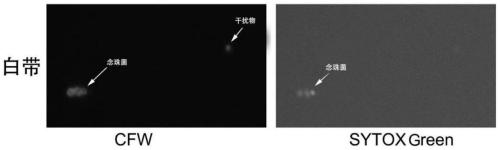
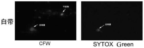
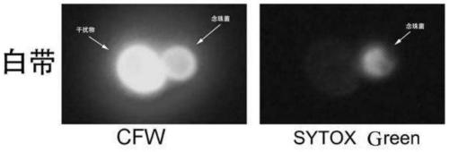
[0039] Disposable leucorrhea swab sampler, professional medical personnel use sterile cotton swabs to collect samples from the upper 1 / 3 section of the side wall of the patient's vagina or the back of the vaginal vault, the cervical canal, etc. Delivered to inspectors within 2 hours.

[0040] Detection process

[0041] 1) Put the sampled cotton swab into the sampler, then add physiological saline, and wash it repeatedly at least submerged in the sample (about 1.5ml), so that the sampling sample is mixed in the physiological saline as much as possible to make a sample suspension;

[0042] 2) Take 500 μl sample suspension, add 1000 μl acetone, shake and mix well, and let it stand for 15 minutes;

[0043]3) Centrifuge at 5000rpm for 10 minutes, suck out the supernatant, add 250 microliters of normal saline to suspend, shake and mix;

[0044] 4) Add 10 μl of SYTOX Green and 125 μl of CFW dye in turn, mix well, and directly use a sampler to draw about 20 μl of liquid and drop it ...


PUM


Abstract

The invention discloses a fungal detection kit comprising a fluorescent brightener, a living cell stain, a fixative, glycerin and NaCl. The fungal detection kit is used for detecting fungi in body fluids; for detecting candida and filamentous bacteria; for detecting leucorrhea and for assisting detection of infectious diseases in the lungs, urinary system and chest and abdomen. The fluorescent brightener dye is used to specifically stain chitin and cellulose of a cell wall of leucorrhea, and the morphology of mold spores and hyphae can be clearly observed under a fluorescence microscope, whileepithelial cells or blood cells from a patient are not pigmented; on the other hand, nucleus staining is performed by the living cell stain, and it is confirmed that the morphology is a cell morphology, thereby rapidly achieving efficient identification of the mold.

Description

technical field [0001] The present application relates to the field of fungal infection detection, in particular to a fluorescent staining detection kit for leukorrhea mold and its application. Background technique [0002] In recent years, the incidence of fungal vaginitis has been on the rise, and it has replaced trichomoniasis as one of the main reasons for the abnormal increase of leucorrhea, which seriously threatens women's health. At present, clinical laboratory diagnosis of fungal vaginitis still mainly relies on direct smear microscopy to find evidence of fungal spores and hyphae infection under a common microscope, but the disadvantage is that the positive rate is too low, and a high proportion of patients with clinical symptoms conform to Fungal infection but microscopic examination was negative. The main defects of conventional microscopic examination are: 1) The morphology of spores or hyphae under the ordinary microscope is easily confused with the same oval w...

Claims


Application Information

Patent Timeline
Patent Type & Authority Applications(China)
IPC IPC(8): C12Q1/04C12R1/72C12R1/645
CPCC12Q1/04G01N2333/37G01N2333/40
Inventor 沈弢张浩吕琳婷郑佳佳许强屠静王誉雅
Owner PEKING UNIV
Why Eureka
Try Eureka