Blood vessel segmentation method, device, medical imaging equipment and storage medium
A blood vessel image and blood vessel technology, applied in neural learning methods, image analysis, image enhancement and other directions, can solve the problems of inability to distinguish coronary structure and background area well, low blood vessel ratio, etc., to improve the accuracy of blood vessel segmentation. Effect
- Summary
- Abstract
- Description
- Claims
- Application Information
AI Technical Summary
Problems solved by technology
Method used
Image


Examples
Embodiment 1
[0034] figure 1 It is a flow chart of a blood vessel segmentation method in Embodiment 1 of the present invention, and this embodiment of the present invention is applicable to the situation of separating blood vessel tissues from background regions in a blood vessel image. The method is executed by a blood vessel segmentation device, which is implemented by software and / or hardware, and is specifically configured in medical imaging equipment. Wherein, the medical imaging equipment may be CT (Computed Tomography, computerized tomography) equipment, MRI (Magnetic Resonance Imaging, magnetic resonance imaging) equipment, DSA (Digital Subtraction Angiography, digital subtraction angiography) equipment, etc.
[0035] Such as figure 1 A blood vessel segmentation method shown includes:
[0036] S110. Acquire a blood vessel image to be segmented and a blood vessel distribution map corresponding to the blood vessel image to be segmented.
[0037] Wherein, the blood vessel image to ...
Embodiment 2
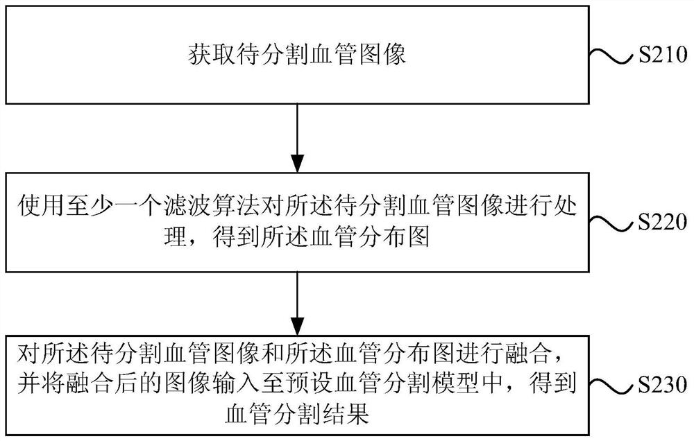
[0047] figure 2 It is a flowchart of a blood vessel segmentation method in Embodiment 2 of the present invention. The embodiment of the present invention performs subdivision optimization on the basis of the technical solutions of the above-mentioned embodiments.
[0048] Further, the operation "inputting the vessel image to be segmented and the vessel distribution map into a preset vessel segmentation model" is refined to "fuse the vessel image to be segmented and the vessel map, and fuse the Input the final image into the preset blood vessel segmentation model” to improve the use mechanism of the preset blood vessel segmentation model.
[0049] Further, the operation "obtaining the blood vessel distribution map corresponding to the to-be-segmented blood vessel image" is refined into "using at least one filter algorithm to process the to-be-segmented blood vessel image to obtain the blood vessel distribution map", so as to improve the blood vessel Determination mechanism of...
Embodiment 3
[0066] Figure 3A It is a flow chart of a blood vessel segmentation method in Embodiment 3 of the present invention. The embodiment of the present invention performs additional optimization and subdivision optimization on the basis of the technical solutions of the above-mentioned embodiments.
[0067] Further, before the operation "input the image of the blood vessel to be segmented and the distribution map of blood vessels into the preset blood vessel segmentation model to obtain the result of blood vessel segmentation", add "training the blood vessel segmentation model"; "Training the blood vessel segmentation model" is refined to "according to at least one historical blood vessel image, determine the corresponding historical blood vessel distribution map; input the historical blood vessel image and the historical blood vessel distribution map into the blood vessel segmentation model to be trained, and obtain the same The current blood vessel segmentation results correspond...
PUM
 Abstract
Description
Claims
Application Information
 - R&D
- Intellectual Property
- Life Sciences
- Materials
- Tech Scout
- Unparalleled Data Quality
- Higher Quality Content
- 60% Fewer Hallucinations
Browse by: Latest US Patents, China's latest patents, Technical Efficacy Thesaurus, Application Domain, Technology Topic, Popular Technical Reports.
© 2025 PatSnap. All rights reserved.Legal|Privacy policy|Modern Slavery Act Transparency Statement|Sitemap|About US| Contact US: help@patsnap.com