Comprehensive oral examination device special for stomatology department

An inspection device and stomatological technology, applied in oral mirrors, applications, medical science, etc., can solve the problems of inconvenient observation and use by doctors, non-adjustable downward pressure, inconvenient oral support, etc., and achieve the effect of easy observation and use

Inactive Publication Date: 2019-04-19
秦洪均
View PDF0 Cites 4 Cited by
  • Summary
  • Abstract
  • Description
  • Claims
  • Application Information

AI Technical Summary

Problems solved by technology

[0002] The oral cavity is a very important structure in the human body. With the continuous updating of medical equipment, intraoral endoscopes are more and more widely used in major hospitals and outpatient clinics. Oral endoscopes are specially constructed camera lenses that can extend into the oral cavity. , display clear and magnified images on the computer or TV screen. With the assistance of the endoscope system, the dentist can better discover the lesions on the soft and hard tissues, and allow the patient to understand their own oral teeth intuitively and comprehensively. Surface stains, plaque, dental calculus, gingivitis and other dental diseases and tooth defects that cannot be seen by oneself at ordinary times, and these images can also be stored in the patient's database, together with text, graphics, X-ray images and other information Together, they form a new generation of electronic medical records, which can be easily accessed and used in consultations, academic reports, etc., but the existing dental examination devices for stomatology are still inconvenient to support the patient's oral cavity, which is inconvenient Examination of the patient's oral cavity with magnified lighting, while the tongue depressor has limited downward pressure on the patient's tongue and cannot be adjusted, blocking the doctor's line of sight and making it difficult for the doctor to observe and use

Method used


Image

  • Comprehensive oral examination device special for stomatology department
  • Comprehensive oral examination device special for stomatology department

Examples


Embodiment Construction

[0013] The following will clearly and completely describe the technical solutions in the embodiments of the present invention with reference to the accompanying drawings in the embodiments of the present invention. Obviously, the described embodiments are only some, not all, embodiments of the present invention. Based on the embodiments of the present invention, all other embodiments obtained by persons of ordinary skill in the art without making creative efforts belong to the protection scope of the present invention.

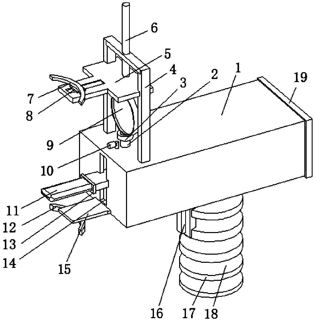
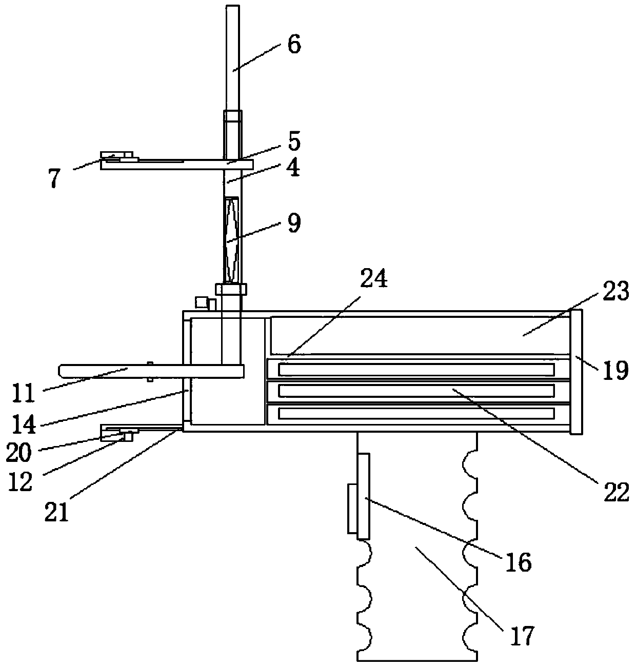
[0014] see Figure 1-2 , the present invention provides a technical solution: a comprehensive oral examination device for stomatology, including a main body shell 1, the inner side of the main body shell 1 is provided with a partition 24, which can support the placement of the battery 23 and the tool 22, the main body shell The left side of 1 is provided with rectangular through groove 14, is positioned at rectangular through groove 14 below and is provided wi...


PUM


Abstract

The invention discloses a comprehensive oral examination device special for the stomatology department. The device comprises a main body shell, a partition plate is arranged on the inner side face ofthe main body shell, a rectangular through groove is formed in the left side of the main body shell, and a lower supporting plate is located on the portion, below the rectangular through groove, on the left side of the main body shell; a second sliding groove is formed in the bottom side of the lower supporting plate, and a second sliding block is slidably connected to the second sliding groove; the bottom side of the second sliding block is provided with an arc-shaped rubber lower brace; a first positioning column is arranged on the portion, on the right side of the arc-shaped rubber lower brace, on the bottom side of the second sliding block; a U-shaped support is arranged at the left end of the upper surface of the main body shell, and an upper supporting plate is slidably connected tothe front side face and the rear side face of the interior of the U-shaped support; a first sliding groove is formed in the upper surface of the upper supporting plate, and a first sliding block is slidably connected to the first sliding groove; the oral cavity of the patient can be well supported, and the oral cavity of the patient can be subjected to amplification, illumination and inspection; pressing force on the tongue of the patient can be adjusted by adjusting a tongue depressor, and observation and application are facilitated for doctors.

Description

technical field [0001] The invention relates to the technical field of cavity inspection devices, in particular to a comprehensive oral cavity inspection device dedicated to stomatology. Background technique [0002] The oral cavity is a very important structure in the human body. With the continuous updating of medical equipment, intraoral endoscopes are more and more widely used in major hospitals and outpatient clinics. Oral endoscopes are specially constructed camera lenses that can extend into the oral cavity. , display clear and magnified images on the computer or TV screen. With the assistance of the endoscope system, the dentist can better discover the lesions on the soft and hard tissues, and allow the patient to understand their own oral teeth intuitively and comprehensively. Surface stains, plaque, dental calculus, gingivitis and other dental diseases and tooth defects that cannot be seen by oneself at ordinary times, and these images can also be stored in the pat...

Claims


Application Information

Patent Timeline
IPC IPC(8): A61B1/24A61B1/06
CPCA61B1/24A61B1/06
Inventor 秦洪均
Owner 秦洪均
Why Eureka
Try Eureka