A method and system for image segmentation of intracranial arteries

An arterial blood vessel and image segmentation technology, applied in the field of medical imaging, can solve the problems of large noise interference and low segmentation operation efficiency, and achieve the effect of small segmentation noise interference and high segmentation operation efficiency.

Active Publication Date: 2021-11-30
UNION STRONG (BEIJING) TECH CO LTD
View PDF6 Cites 0 Cited by
  • Summary
  • Abstract
  • Description
  • Claims
  • Application Information

AI Technical Summary

Problems solved by technology

[0004] However, in the prior art, when the intracranial artery image segmentation method is used for segmentation, the noise interference is large and the efficiency of the segmentation operation is low.

Method used


Image

  • A method and system for image segmentation of intracranial arteries
  • A method and system for image segmentation of intracranial arteries
  • A method and system for image segmentation of intracranial arteries

Examples


Embodiment Construction

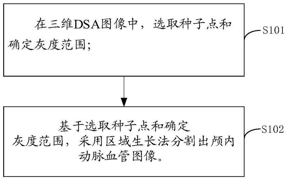
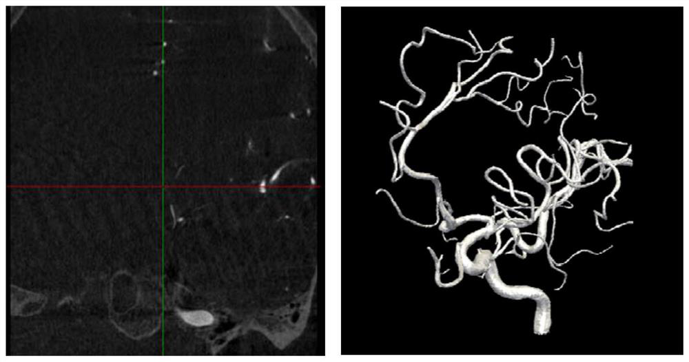
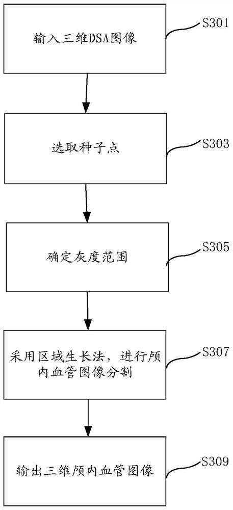
[0026] The embodiment of the present application provides a method and system for image segmentation of intracranial arteries, which are used to solve the problems of large noise interference and low operation efficiency in DSA image segmentation, thereby effectively reducing noise interference in DSA image segmentation and improving operation efficiency.

[0027] In order to make the purpose, technical solution and advantages of the present application clearer, the technical solution of the present application will be clearly and completely described below in conjunction with specific embodiments of the present application and corresponding drawings. Apparently, the described embodiments are only some of the embodiments of the present application, rather than all the embodiments. Based on the embodiments in this application, all other embodiments obtained by persons of ordinary skill in the art without making creative efforts belong to the scope of protection of this applicati...


PUM


Abstract

The embodiment of the present application discloses a method and system for segmenting an intracranial artery image. The scheme includes: selecting seed points and determining the gray scale range in the three-dimensional DSA image; based on the selected seed points and determining the gray scale range, segmenting the intracranial artery image by using the region growing method. This scheme provides a segmentation method of intracranial arterial blood vessel image with less interference of segmentation noise and higher efficiency of segmentation operation.

Description

technical field [0001] The present application relates to the field of medical imaging, in particular to a method and system for segmenting images of intracranial arteries. Background technique [0002] With the continuous progress and development of modern medical imaging equipment, the development of medical imaging technology and computer graphics has brought great changes to medicine. Computed tomography (CT), nuclear magnetic resonance (MRI), ultrasound (US), positron Medical imaging techniques such as emission computed tomography (PET) and digital subtraction angiography (Digital Subtraction Angiography, DSA) have been widely used in clinical diagnosis. DSA is a new X-ray imaging system. It is the product of the combination of conventional angiography and computer image processing technology. It can also be said that the image of bone and soft tissue on the angiogram is eliminated by computer. A photographic technique that highlights blood vessels. [0003] The basic...

Claims


Application Information

Patent Timeline
Patent Type & Authority Patents(China)
IPC IPC(8): G06T7/11G06T7/136
CPCG06T2207/10081G06T2207/10104G06T2207/30016G06T2207/30096G06T2207/30101G06T7/11G06T7/136
Inventor 杨光明冯雪王文智秦岚
Owner UNION STRONG (BEIJING) TECH CO LTD
Why Eureka
Try Eureka