Method of color normalization of pathological images based on low-rank embedded non-negative matrix factorization

A non-negative matrix decomposition, pathological image technology, applied in image analysis, image enhancement, medical image and other directions, can solve the problems of large color difference of pathological images, the influence of intelligent analysis, image color inconsistency, etc., to achieve simple algorithm, efficient color standardization , the effect of fast calculation

Active Publication Date: 2021-09-17
HEFEI UNIV OF TECH
View PDF2 Cites 0 Cited by
  • Summary
  • Abstract
  • Description
  • Claims
  • Application Information

AI Technical Summary

Problems solved by technology

Due to the differences in the scanning methods of different digital slide scanners, different laboratory staining methods and the ratio of dyeing agents, there are large color differences in pathological images, and image color inconsistencies under the same staining method occur from time to time. Intelligent analysis of both diagnostic and pathological images has an impact

Method used


Image

  • Method of color normalization of pathological images based on low-rank embedded non-negative matrix factorization
  • Method of color normalization of pathological images based on low-rank embedded non-negative matrix factorization
  • Method of color normalization of pathological images based on low-rank embedded non-negative matrix factorization

Examples


Embodiment Construction

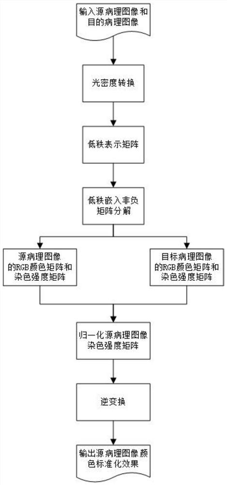
[0042] like figure 1 Shown, based on low rank NMF embedded pathological standardized color image, comprising the following steps: Step (1): Histology Histology Source by the scanner to be converted and as a standard to scan a target Histology computer stores RGB three-channel color image, to obtain a source image and the target pathological pathological image.

[0043] Step (2): the pathological image source image and the target pathological converted to the corresponding optical density of the image.

[0044] Step (3): In the image pixels, the optical density of each pixel is represented as a three-channel three-dimensional vector, i.e. as a sample point for each pixel, each sample point as a three-dimensional vector, whereby the source pathological image and the target image into a pathological form of a matrix, the number of lines is 3, the number of columns is the number of pixels or said number of samples. Respectively, then using low rank indicates a low rank pathological im...


PUM


Abstract

The invention discloses a pathological image color standardization method based on low-rank embedded non-negative matrix decomposition, which expresses two pathological image data in the form of observation matrix, performs optical density conversion on them respectively, and converts the source optical density matrix after conversion and the target optical density matrix to solve the low-rank representation matrix respectively, decompose the corresponding optical density matrix into the RGB color matrix of each dyeing component and the intensity matrix of each pixel under each dyeing component, and finally convert it into the image RGB form to achieve For the purpose of color standardization from source pathological images to target pathological images, the present invention can be used for color standardization of pathological images and improvement of visualization effects, avoiding color inconsistencies in pathological images caused by differences in different pathological scanners, different laboratory dyeing methods and dye ratios It is of great significance to pathological image imaging, dye separation and intelligent pathological image analysis, and has broad market prospects and application value.

Description

Technical field [0001] The present invention relates to digital image processing technology, and particularly relates to a color image normalized digital image processing technology, and particularly relates to a method based on low rank NMF embedded pathological image color standardization. Background technique [0002] Digital pathology image display color information for different structures by a histopathological stain staining, is the most representative hematoxylin - eosin. Since different degrees of differences digital scanner slice method, different laboratories and staining dye ratio, such that there is a large color pathologic image differences have occurred under the same staining method inconsistent color image, the doctor diagnostic and pathological intelligent image analysis are affected. Thus, a color image is needed is a standardized method of pathology, pathology image to avoid affecting the color differences caused by, a pathological image to improve the color q...

Claims


Application Information

Patent Timeline
Patent Type & Authority Patents(China)
IPC IPC(8): G06T7/90G06T5/00G16H30/20
CPCG06T5/005G06T7/90G16H30/20
Inventor 史骏饶诗语郑利平
Owner HEFEI UNIV OF TECH
Why Eureka
Try Eureka