A method for inducing differentiation of amniotic epithelial stem cells into functional liver cells and its application
A technology for inducing differentiation and stem cells, applied in cell dissociation methods, cell culture active agents, biochemical equipment and methods, etc., can solve the problems of difficult clinical practice of cells, tumorigenicity of stem cells, limited sources of bone marrow and umbilical cord blood, etc. Achieve the effect of enriching the source of cells, inhibiting the activity of transaminase, and restoring the level of albumin in the liver
- Summary
- Abstract
- Description
- Claims
- Application Information
AI Technical Summary
Problems solved by technology
Method used
Image


Examples
Embodiment 1
[0089] Example 1. Isolation, culture, expansion and GFP marker of amniotic membrane epithelial stem cells (HAESCs):
[0090] Under the premise of obtaining the consent of the newborn's family members, the fresh amniotic membrane was isolated. After 2 hours of antibiotic treatment, microbiological detection and safety detection of infectious disease pathogens were carried out. After the amniotic membrane was digested by trypsin / EDTA (0.25%) in a water bath at 37°C for 1 hour, stop solution was added. After centrifugation, the supernatant was removed, and the amnion epithelial stem cell culture medium was placed in 5% CO 2 , 95% humidity, and cultured in a 37°C incubator. Change the medium after 2-3 days to remove unattached cells, and then change the medium every 2 days. When the confluence of the cells reached 80%, trypsin / EDTA was used for passage. The amniotic epithelial stem cells of passage 3 were transfected with GFP using lentivirus. Immunofluorescence and flow cyto...
Embodiment 2
[0176] Example 2. In vivo and in vitro tumorigenicity detection of amniotic membrane epithelial stem cells
[0177] 1. In vivo and in vitro tumorigenicity detection of amniotic membrane epithelial stem cells
[0178]1) Take the third-generation amnion epithelial cells in good growth condition, which are characterized by vigorous growth, large cell bodies, clear nuclei, rich cytoplasm, and strong refraction under the microscope, and digest them with trypsin-EDTA (0.25% ) digestion, under the microscope, when the cells became a single round shape, the digestion was terminated with DMEM medium containing 10% (volume concentration) fetal bovine serum, centrifuged after gentle blowing, and the cell pellet was obtained, washed with PBS (PBS without calcium and magnesium ions) solution) and washed twice (the purpose is to wash away trypsin, fetal bovine serum and other substances);
[0179] 2) Inoculate the amnion epithelial stem cells in step 1) on soft agar, and after culturing fo...
Embodiment 3
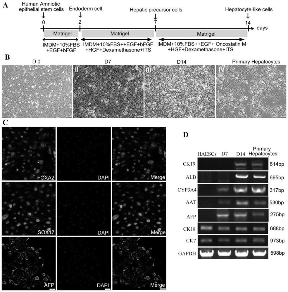
[0187] Example 3. Induced differentiation of amniotic membrane epithelial stem cells into hepatocytes and its function detection
[0188] The specific operating procedures are as follows:
[0189] 1. Directed induction of amniotic epithelial stem cells to differentiate into hepatocytes in vitro
[0190] 1) Take the well-growing cells of the third generation and digest them with trypsin-EDTA digestion solution (0.25%). The DMEM medium was digested, centrifuged after gently pipetting to obtain cell pellets, and washed twice with PBS (PBS washing solution without calcium and magnesium ions) (the purpose is to wash away trypsin, fetal bovine serum and other substances);
[0191] 2) The above-mentioned cell pellets washed with PBS were inoculated into Matrigel pretreated 6-well plates for induction culture, and the seeding cell density was 3×10 5 cells / well, add 2ml of amniotic epithelial stem cell general medium to each well, at 37°C, 5% CO 2 , 95% saturated humidity incubator ...
PUM
 Abstract
Description
Claims
Application Information
 - R&D
- Intellectual Property
- Life Sciences
- Materials
- Tech Scout
- Unparalleled Data Quality
- Higher Quality Content
- 60% Fewer Hallucinations
Browse by: Latest US Patents, China's latest patents, Technical Efficacy Thesaurus, Application Domain, Technology Topic, Popular Technical Reports.
© 2025 PatSnap. All rights reserved.Legal|Privacy policy|Modern Slavery Act Transparency Statement|Sitemap|About US| Contact US: help@patsnap.com