
Ultrasound breast tumor grading method based on multi-feature extraction and Linear SVM

A breast tumor and grading method technology, which is applied in the field of computer-aided diagnosis of medical images, can solve the problem of no related research on the quantitative grading of breast tumors, and achieve the effects of reducing the influence of subjective factors, improving accuracy, and satisfying the time complexity of the algorithm

Inactive Publication Date: 2018-12-21
JIANGSU PROVINCIAL HOSPITAL OF TCM
View PDF7 Cites 5 Cited by
  • Summary
  • Abstract
  • Description
  • Claims
  • Application Information

AI Technical Summary

Problems solved by technology

However, there is no relevant research on the quantitative grading of breast tumors with grades 3, 4, and 5.

Method used


Image

  • Ultrasound breast tumor grading method based on multi-feature extraction and Linear SVM
  • Ultrasound breast tumor grading method based on multi-feature extraction and Linear SVM
  • Ultrasound breast tumor grading method based on multi-feature extraction and Linear SVM

Examples


Embodiment Construction

[0020] The present invention will be further described below in conjunction with accompanying drawing.

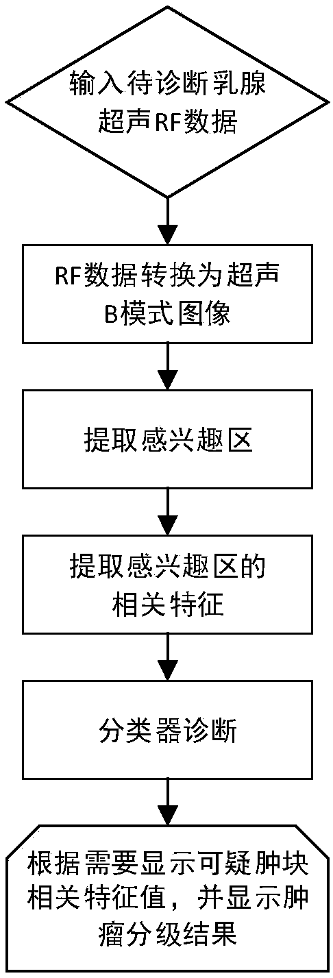
[0021] The overall process of the present invention is divided into three parts: acquiring RF data from ultrasound and performing image segmentation; multi-feature extraction; classification model training and verification. The specific steps are as figure 1 shown, including:

[0022] 1) Input a frame of breast ultrasound RF data to be diagnosed;

[0023] 2) Convert the input breast RF data into an ultrasound B-mode image, and outline the location of the initial suspicious mass area;

[0024] 3) Segment the suspicious mass within the initial suspicious mass region, and determine the boundary of the suspicious mass;

[0025] 4) Calculate the eigenvalues ​​of the segmented suspicious mass regions;

[0026] 5) Input the calculated eigenvalues ​​into the classifier to analyze the suspicious mass area.

[0027] The eigenvalues ​​of the tumor area calculated in step 4) are d...


PUM


Abstract

The invention discloses an ultrasound breast tumor grading method based on multi-feature extraction and Linear SVM. The method comprises steps that to-be-diagnosed breast ultrasound RF data is inputted; the RF data is converted into an ultrasound B-mode image to obtain the initial suspicious lump area location; the initial suspicious lump area is segmented to obtain a suspicious lump to determinethe boundary of the suspicious lump; a feature value of the segmented suspicious lump is calculated; the calculated feature value is inputted into a classifier, and the suspicious lump area is analyzed. The method is advantaged in that based on the BI-RADS grading standard, the actually acquired breast ultrasound RF data is hierarchically detected, breast tumor ultrasound 3, 4 and 5-level classification is achieved through utilizing the multi-feature extraction method and the linear support vector machine classifier, improvement of accuracy of the doctor's diagnosis is facilitated, the algorithm time and complexity can meet clinical requirements, and the method has a certain practical value.

Description

technical field [0001] The invention belongs to the application field of computer-aided diagnosis of medical images, and in particular relates to an ultrasonic mammary gland tumor grading method based on multi-feature extraction and Linear SVM. Background technique [0002] Breast tumor is an extremely common tumor lesion that endangers the health of women. So far, no research can reveal the exact cause of breast tumor. Early diagnosis and grading are crucial for breast tumors. Breast ultrasonography and breast biopsy are the main means of evaluating benign and malignant breast tumors. Although breast biopsy is the gold standard for tumor evaluation, biopsy is invasive, expensive, and extremely uncomfortable for patients. In addition, many biopsy results are benign. Therefore, there is a need for improved ultrasound diagnosis to reduce unnecessary biopsies. Ultrasound diagnosis of the breast mainly comes from the qualitative evaluation of the breast tissue and the soft tiss...

Claims


Application Information

Patent Timeline
Patent Type & Authority Applications(China)
IPC IPC(8): G16H50/20G06T7/11G06T7/12G06K9/62
CPCG06T7/11G06T7/12G16H50/20G06T2207/20104G06T2207/30096G06T2207/30068G06T2207/10132G06T2207/20081G06F18/2411
Inventor 严郁吴意赟朱伟蔡晓巍蔡润秋蒋文婧
Owner JIANGSU PROVINCIAL HOSPITAL OF TCM
Learn More