A Method for Analyzing Anterior Segment Morphology

A technology of morphological analysis and anterior segment, applied in gonioscopy, eye testing equipment, medical science, etc., can solve the problem of inability to quantitatively analyze the morphological parameters of the anterior segment, achieve easy acceptance, ensure measurement accuracy, and reduce algorithm complexity degree of effect

Active Publication Date: 2020-10-16
TIANJIN SUOWEI ELECTRONICS TECH
View PDF6 Cites 0 Cited by
  • Summary
  • Abstract
  • Description
  • Claims
  • Application Information

AI Technical Summary

Problems solved by technology

The system can only perform image observation, but cannot quantitatively analyze the morphological parameters of the anterior segment

Method used


Image

  • A Method for Analyzing Anterior Segment Morphology
  • A Method for Analyzing Anterior Segment Morphology
  • A Method for Analyzing Anterior Segment Morphology

Examples


Embodiment Construction

[0031] The implementation of the present invention will be described in further detail below in conjunction with the accompanying drawings. The following embodiments are only descriptive, not restrictive, and cannot limit the protection scope of the present invention.

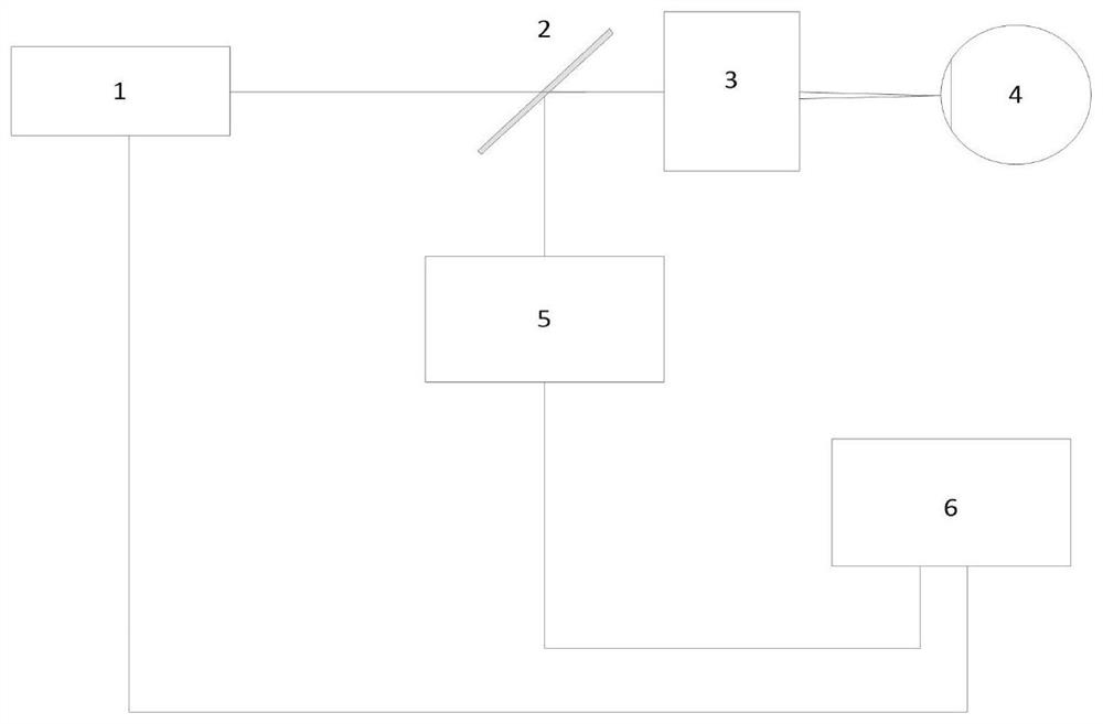
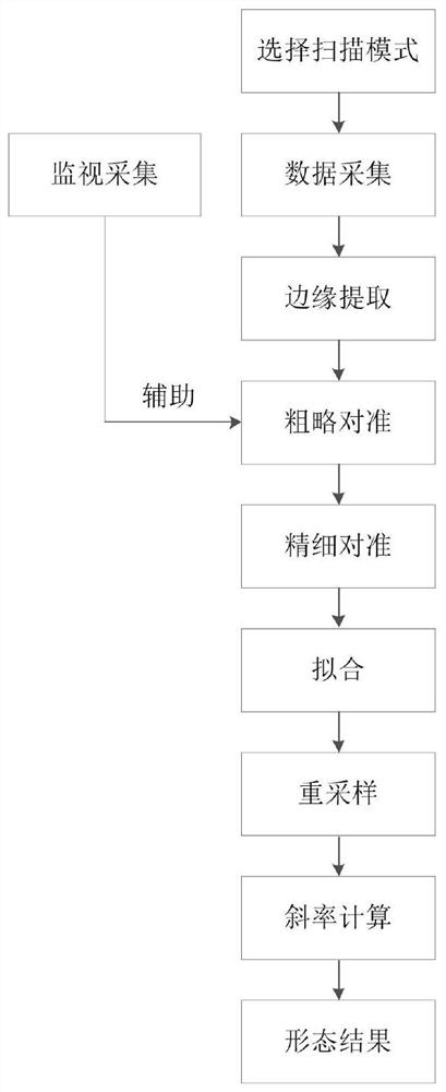
[0032] An anterior segment morphology analysis method, such as figure 1 As shown, the data acquisition system adopted in this method includes a monitoring module 1, a spectroscopic module 2, an eyepiece 3, an optical coherence tomography module 5 and a computer 6, and performs two-dimensional vibrating mirror non-contact optical scanning on the human eye 4 to collect Section three-dimensional data blocks, such as figure 2 As shown, the method includes the following steps:

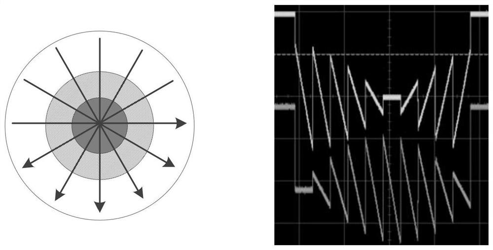
[0033] (1) Select the scanning mode, the scanning mode adopts rotary scanning, the driving signal of the two-dimensional vibrating mirror used is: both directions are driven by sawtooth wave, and the one-to-one correspondence in timing is ...


PUM


Abstract

The invention relates to an anterior segment morphology analysis method, wherein the data acquisition system used by the method comprises a monitoring module, a light splitting module, an eyepiece, anoptical coherence tomography module and a computer. A two-dimensional galvanometer non-contact optical scan is performed on a human eye to collect a three-dimensional data block of the anterior segment of the eye, and the method comprises the following steps of: acquiring the three-dimensional data block of the anterior segment, performing edge extraction on the data block, carrying out rough alignment and fine alignment, fitting, re-sampling and calculating a slope, and finally obtaining the tissue morphology analysis result of the anterior segment, including an anterior segment sectional image, a corneal anterior surface topographic map, a corneal posterior surface topographic map, a corneal thickness map, a crystalline anterior surface topographic map, a crystalline posterior surface topographic map, a crystalline thickness map, a corneal three-dimensional image, a chamber-angle three-dimensional image, an iris three-dimensional image, and a crystalline three-dimensional image. Byusing the method, the complexity of the algorithm is reduced, the measurement accuracy is ensured. The method is non-invasive, has no stimulation, and is easily accepted by patients.

Description

technical field [0001] The invention belongs to the technical field of ophthalmic medical equipment, and relates to an anterior segment shape analysis method. Background technique [0002] In the concept of medical eye anatomy, the anterior segment includes the part of the eye tissue from the cornea to the lens. In recent years, the prevalence of the anterior segment has been increasing year by year, so the examination of the cornea is particularly important. At the same time, the condition of the lens is also an important reference in the diagnosis of glaucoma, cataract and refractive vision problems. [0003] Traditional anterior segment imaging techniques can generally observe the anterior segment from the cornea to the front surface of the lens, but cannot cover the entire lens area. In terms of anterior segment morphology analysis, corneal topography based on the Pacido disc principle is currently one of the most widely used techniques. This technique is easily affect...

Claims


Application Information

Patent Timeline
Patent Type & Authority Patents(China)
IPC IPC(8): A61B3/117A61B3/14
CPCA61B3/117A61B3/14
Inventor 赵鹏王雪乔孟庆宾张雪峰王元张锟李朋勃齐岳
Owner TIANJIN SUOWEI ELECTRONICS TECH
Why Eureka
Try Eureka