
Hemostasis device for medical gynecological surgery

A technology for surgery and obstetrics and gynecology, which is applied in the field of hemostatic devices for medical obstetrics and gynecology surgery, which can solve the problems of affecting the hemostasis effect and increasing the pain of patients, and achieve the effects of improving hemostasis efficiency, improving treatment efficiency, and reducing pain

Active Publication Date: 2018-10-19
NANYANG CITY CENT HOSPITAL
View PDF7 Cites 2 Cited by
  • Summary
  • Abstract
  • Description
  • Claims
  • Application Information

AI Technical Summary

Problems solved by technology

[0004] The purpose of the present invention is to provide a hemostatic device for medical obstetrics and gynecology surgery, which solves the problem that the hemostatic balloon moves in the uterus, affects the hemostatic effect, and increases the pain of the patient

Method used


Image

  • Hemostasis device for medical gynecological surgery
  • Hemostasis device for medical gynecological surgery
  • Hemostasis device for medical gynecological surgery

Examples


Embodiment Construction

[0024] The specific embodiments of the present invention will be described in detail below in conjunction with the accompanying drawings, but it should be understood that the protection scope of the present invention is not limited by the specific embodiments.

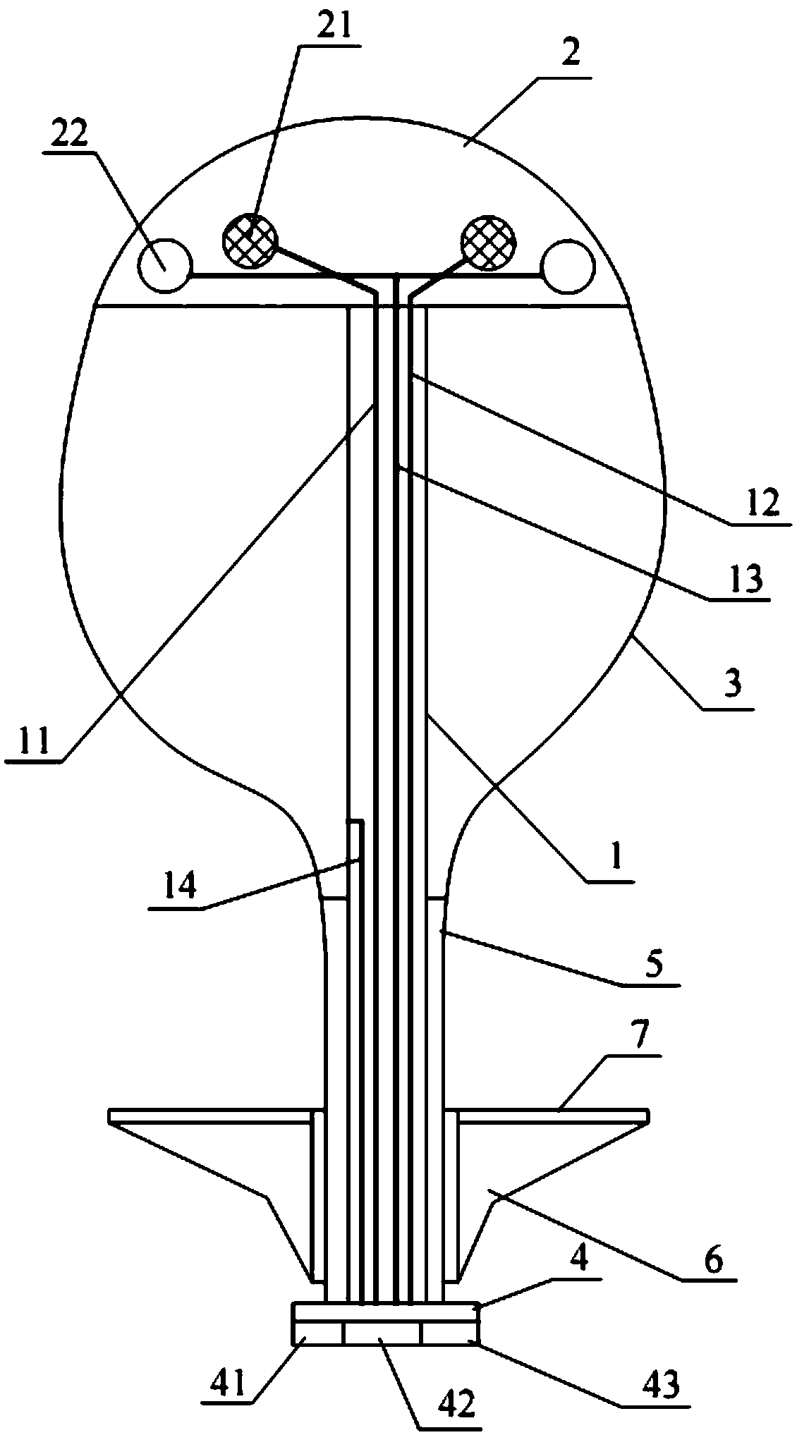
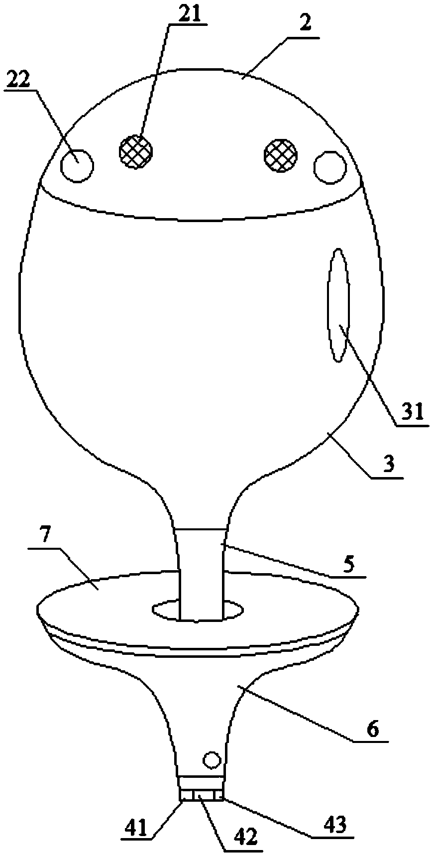
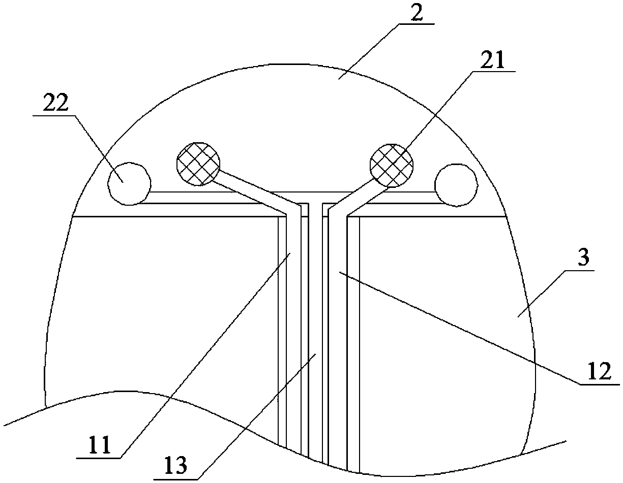
[0025] A hemostatic device for medical obstetrics and gynecology operations, specifically as Figure 1-6 As shown, it includes the whole body, the whole body includes a channel pipe 1 and a hollow spherical support body 2, the height of the support body 2 is less than its spherical radius, the support body 2 is installed at the far end of the channel pipe 1 and communicates with the channel pipe 1, that is The support body 2 is installed on the end of the channel tube 1 away from the operator's hand, and the channel tube 1 is sequentially sleeved with the main capsule body 3 and the fixing assembly from the distal end to the proximal end, and the main capsule body 3 is in contact with the plane of the support body 2 Co...


PUM


Abstract

The invention belongs to the technical field of medical hemostasis devices, and particularly relates to a hemostasis device for a medical gynecological surgery. The device comprises a through body, the through body comprises a channel tube and a hollow segment-shaped support body, the support body is installed in the distal end of the channel tube and communicated with the channel tube, a capsulebody and a fixing assembly sleeve from the distal end to the proximal end of the channel tube, the main capsule body is in contact with the support body, and a sealing plate is further mounted on theproximal end of the channel tube; an absorption net and a suction cup are mounted on the spherical surface of the support body; the fixing assembly includes a fixing sleeve, a sub-capsule body and a contact plate. The device achieves the fixation of a hemostatic ball capsule by arranging the suction cup and the fixing assembly on the support body, and capsule body compression hemostasis and medicine hemostasis are simultaneously performed, thereby improving the hemostatic efficiency, reducing the pain of a patient and adsorbing pollutants in the uterus.

Description

technical field [0001] The invention belongs to the technical field of medical hemostatic devices, in particular to a medical hemostatic device for obstetrics and gynecology operations. Background technique [0002] After giving birth, some women will experience postpartum hemorrhage, especially those with placenta previa. Because the placenta is located in the lower uterine segment and the internal opening of the cervix, it often leads to massive bleeding. If the bleeding is not stopped in time, it will endanger the life of the mother in severe cases. Although hemostatic drugs can promote uterine contractions and effectively reduce bleeding, the drugs need to undergo blood absorption and chemical reactions to have a better hemostatic effect. Filling the uterus with gauze to stop bleeding is an effective and rapid method of hemostasis, but filling the gauze is a very primitive and simple means of hemostasis. The surface of the gauze is rough and smooth. environment, increas...

Claims


Application Information

Patent Timeline
IPC IPC(8): A61B17/42A61B17/12A61M31/00
CPCA61B17/12A61B17/4241A61B2017/12004A61B2017/4216A61M31/00A61M2210/1433A61M2210/005
Inventor 马万增欧阳晟姚鑫
Owner NANYANG CITY CENT HOSPITAL
Learn More