
Design method of 3D printing template for intracranial tumor positioning, drilling and puncturing operations

A tumor localization and 3D printing technology, which is applied in the field of clinical medicine, can solve the problems of poor treatment effect, great patient pain, and low accuracy, and achieve the effects of improved work efficiency, simple operation, and reduced cost

Pending Publication Date: 2018-09-14
李成利 +1
View PDF7 Cites 6 Cited by
  • Summary
  • Abstract
  • Description
  • Claims
  • Application Information

AI Technical Summary

Problems solved by technology

[0005] The present invention aims at the technical defects of the prior art, and provides a design method of a 3D printing template for brain tumor positioning, drilling, and puncture operations, so as to solve the problem that such operations in the prior art are mainly performed by doctors based on experience. There are technical problems such as low accuracy, poor treatment effect, large error, long time, and great pain for patients

Method used


Image

  • Design method of 3D printing template for intracranial tumor positioning, drilling and puncturing operations

Examples


Embodiment 1

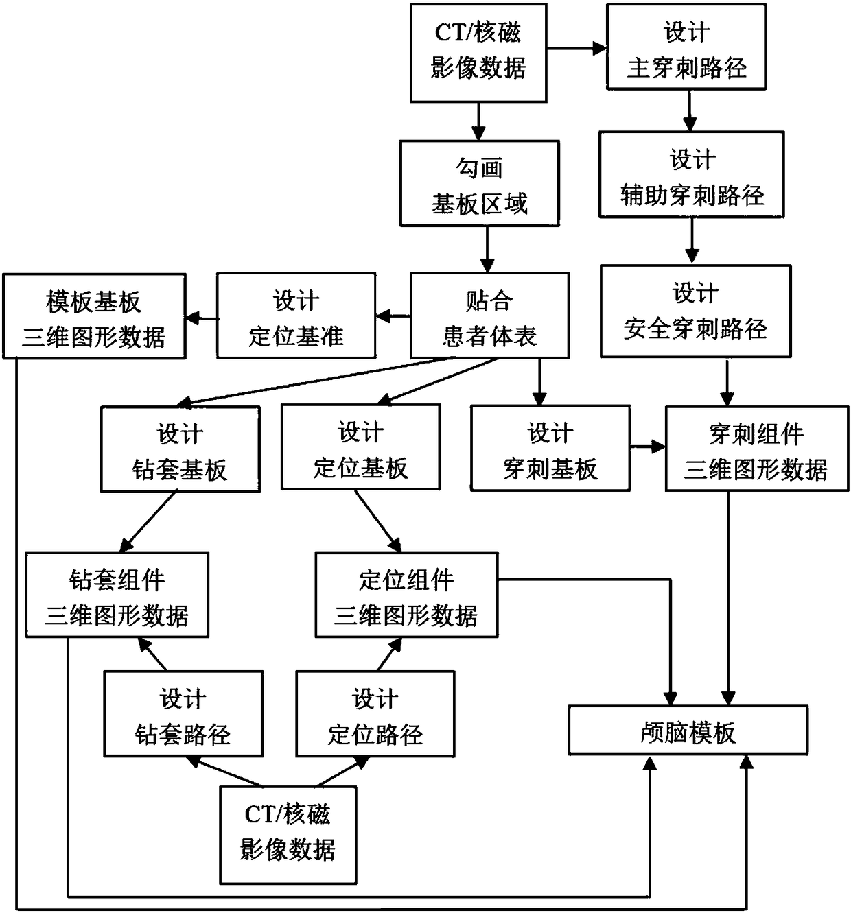
[0027] Design method of 3D printing template for craniocerebral tumor localization, drilling and puncture. This method designs and produces craniocerebral templates according to the needs of patients' individualized precision medicine, the shape of brain tumors and the relationship with surrounding biological tissues It consists of two parts: replaceable components and template substrate. The replaceable assemblies are the positioning assembly, the drill sleeve assembly and the piercing assembly, respectively.

[0028] 1. Design positioning components

[0029] On the special positioning component design software, the two-point positioning method is used to select a safe and reasonable skull drilling point and the best path for the brain tumor target area (hereinafter referred to as the target area) as the center line, and avoid important brain tissue. The functional area, blood vessels, nerves and other tissues are designed as a hollow cylinder with a diameter of 3 to 10 mm (...

Embodiment 2

[0049] The design method of 3D printing template for craniocerebral tumor localization, drilling and puncture operation, including the following steps:

[0050] 1) Design the positioning component: On the positioning component design software, the path from the skull drilling point to the brain tumor target area is determined by the two-point positioning method, and the path avoids the functional area of ​​the brain tissue, blood vessels, and nerve tissue; the design diameter is 3 ~10mm, 15mm high, 0.5~1mm thick hollow cylinder, filled with cod liver oil;

[0051] 2) Design the drill sleeve assembly: On the drill sleeve assembly design software, determine the T-shaped drill sleeve for the bone drill according to the path and size described in step 1). The wall thickness of the T-shaped drill sleeve is 2-3 mm, The inner diameter of the T-shaped drill sleeve is 0.2 mm larger than the diameter of the drill bit of the T-shaped bone drill, the T-shaped drill sleeve is made of titan...


PUM

PropertyMeasurementUnit
Diameteraaaaaaaaaa
Wall thicknessaaaaaaaaaa
Thicknessaaaaaaaaaa

Abstract

The invention provides a design method of a 3D printing template for intracranial tumor positioning, drilling and puncturing operations. The method comprises the steps of design of a replaceable assembly and design of a template substrate, wherein the replaceable assembly specifically comprises a positioning assembly, a drill bush assembly and a puncturing assembly. The method provided by the invention takes the position and the form of a brain tumor of a patient and the relation of the brain tumor with surrounding tissues as a basis, generates a three-dimensional data model based on a software tool, and can be directly used for executing 3D printing. The product configuration obtained by printing can accurately reflect the true spatial configuration of a lesion area and can completely restore the design idea of a clinician, so that the safety of a puncturing operation and the treatment effect are ensured, the working efficiency is greatly improved, the cost is obviously lowered, the operation is simple, and the maintenance is convenient. Furthermore, because the method can adapt to specific conditions of various cases, the requirements of individualized and precise medical treatment for patients can be met.

Description

technical field [0001] The invention relates to the technical field of clinical medicine, as well as medical equipment and 3D printing technology, in particular to a design method of a 3D printing template for craniocerebral tumor localization, drilling and puncture operations. Background technique [0002] Puncture surgery is a clinically necessary technique for tumor biopsy and further treatment, and is used to provide the basis for the pathological diagnosis of tumors. Brain tumors, also known as intracranial tumors, refer to tumors of the nervous system that occur in the cranial cavity, including tumors originating from neuroepithelium, peripheral nerves, meninges and germ cells, tumors of lymphoid and hematopoietic tissues, and craniopharyngiomas in the sella region with granulosa cell tumors, as well as metastatic tumors. With the continuous improvement of inspection methods and methods, the accuracy of diagnosis has gradually increased, but there are still a large nu...

Claims


Application Information

Patent Timeline
IPC IPC(8): A61B34/20A61B17/17A61B17/34B33Y80/00
CPCA61B17/17A61B17/1703A61B17/3403A61B34/20B33Y80/00A61B2034/2068
Inventor 李成利姜冠群谢大鹏
Owner 李成利
Learn More