Ophthalmological function checking device based on slit lamp platform and image processing method
A functional examination, slit lamp technology, applied in image data processing, image enhancement, image analysis, etc., can solve the problems of lack of non-invasive microvascular function evaluation and analysis methods, restricting the progress of ocular surface microvascular research, etc.
- Summary
- Abstract
- Description
- Claims
- Application Information
AI Technical Summary
Problems solved by technology
Method used
Image

Examples
Embodiment 1
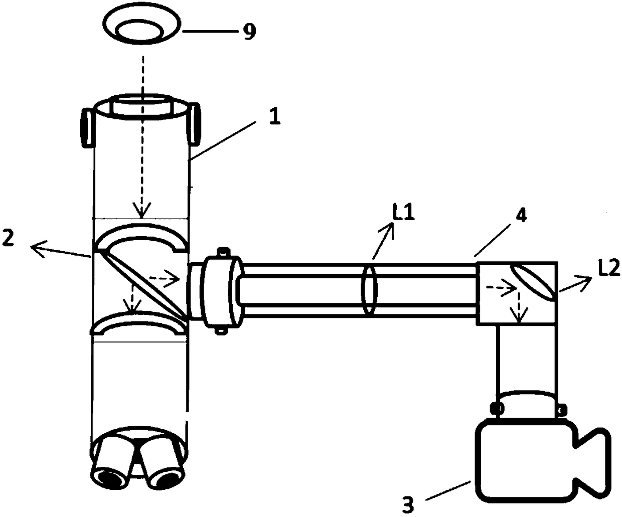
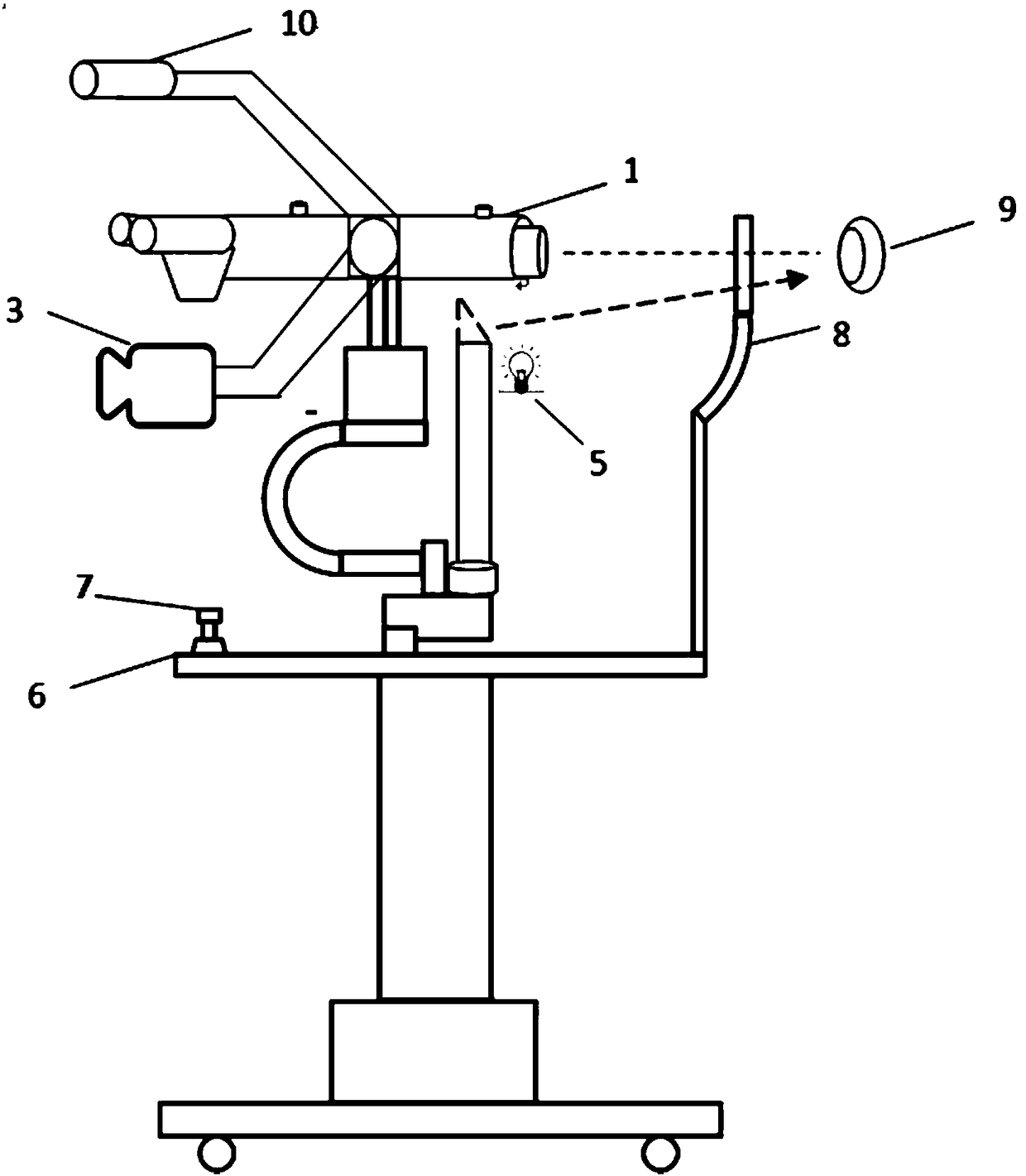
[0040] Such as figure 1 and 2 As shown, an ophthalmic function inspection device based on a slit lamp platform includes a slit lamp lifting platform 6, an imaging device supported on the slit lamp lifting platform 6, and the imaging device includes a slit lamp microscope 1, an optical path lens barrel 4 and A high-speed camera 3 connected to the slit lamp microscope 1, the slit lamp microscope 1 can observe the light reflected by the eyeball, the light reflected by the eyeball enters the high-speed camera 3 through the optical path lens barrel 4, and the high-speed camera 3 can form an original image from the light reflected by the eyeball , because the light reflected by the eyeball contains the information of the microvessels on the surface of the eyeball, and the original image contains the information of the microvessels of the eyeball. The slit lamp, the lifting principle of the slit lamp elevating platform 6, the slit lamp microscope 1 and the high-speed camera 3 all ad...
Embodiment 2
[0050] This embodiment is to perform complexity analysis on the eye microvessel image acquired in the first embodiment.
[0051] The method for analyzing the complexity of eye microvessels in the image comprises the following steps:
[0052]Step 1: Preprocessing the original image acquired by the high-speed camera 3 .
[0053] It specifically includes the following steps: step a): removing Gaussian noise and salt-and-pepper noise in the image, specifically, moving the Gaussian filter to filter the Gaussian noise first, and then removing the salt-and-pepper noise through median filtering. The principles of the Gaussian filter and the median filter belong to the prior art and will not be described in detail here.
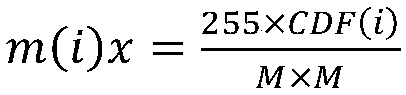
[0054] Step b): The image contrast is enhanced by Contrast-Limited Adaptive Histogram Equalization (CLAHE) on the image after denoising. Contrast enhancement can be defined as the slope of the grayscale mapping function. When the sliding window size is set to MxM, t...
Embodiment 3
[0080] This embodiment calculates the diameter of the microvessels on the surface of the eyeball. When a lesion occurs in the eye, the diameter of the eye capillary will change. Therefore, by calculating the diameter of the eye microvessel, it is possible to determine whether the eye has a lesion and the degree of the lesion.
[0081] Before calculating the diameter of the eye microvessels, it is necessary to preprocess the eye microvessel images acquired by the high-speed camera 3. The preprocessing process is the same as that in Embodiment 2, and will not be described in detail here.
[0082] When calculating the diameter, the centerline of the eye microvessels was obtained by using the centerline extraction method based on the Hessian matrix, and then the width of the eye microvessels perpendicular to the center line was calculated as the diameter of the eye microvessels.
[0083] Let (xi, yi) be any point on the center line, and use the multi-point fitting on the center lin...
PUM
 Abstract
Description
Claims
Application Information
 - R&D
- Intellectual Property
- Life Sciences
- Materials
- Tech Scout
- Unparalleled Data Quality
- Higher Quality Content
- 60% Fewer Hallucinations
Browse by: Latest US Patents, China's latest patents, Technical Efficacy Thesaurus, Application Domain, Technology Topic, Popular Technical Reports.
© 2025 PatSnap. All rights reserved.Legal|Privacy policy|Modern Slavery Act Transparency Statement|Sitemap|About US| Contact US: help@patsnap.com