Fetus panorama display instrument based on ultrasonic and 3D technology

A fetal and panoramic technology, applied in the field of fetal panoramic display based on ultrasound and 3D technology, can solve the problems of insufficient affinity and intuitiveness, poor human-computer interaction effect, and unfriendly interface.

Inactive Publication Date: 2018-06-22
FOURTH MILITARY MEDICAL UNIVERSITY
View PDF4 Cites 3 Cited by
  • Summary
  • Abstract
  • Description
  • Claims
  • Application Information

AI Technical Summary

Problems solved by technology

Not friendly and intuitive enough for parents to construct the concept of the overall image of the fetus
Most of the models established by 3D B-ultrasound scanning are operated by clicking and dragging the mouse on the screen to rotate and other operations. The human-computer interaction effect is relatively poor, and the interactive interface is relatively unfriendly.

Method used


Image

  • Fetus panorama display instrument based on ultrasonic and 3D technology
  • Fetus panorama display instrument based on ultrasonic and 3D technology
  • Fetus panorama display instrument based on ultrasonic and 3D technology

Examples


specific Embodiment approach

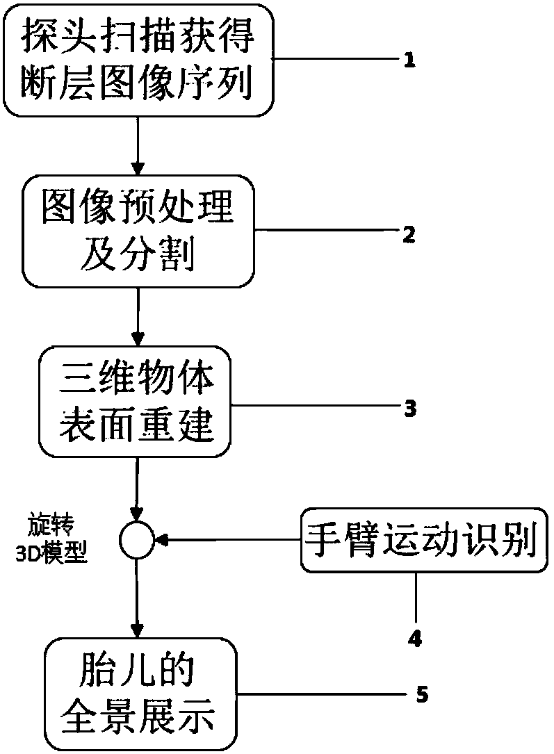
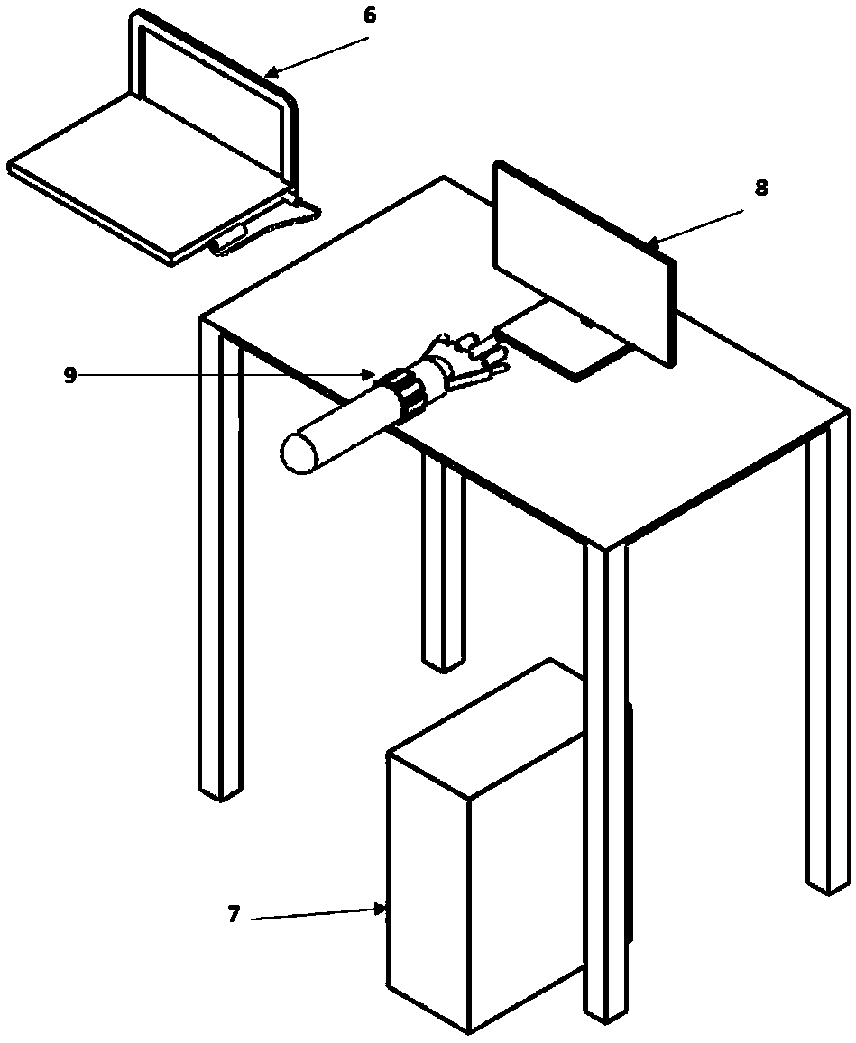
[0030] 1) Use the B-ultrasound 6 probe to scan to obtain tomographic image sequence, or directly obtain the fetal ultrasound image sequence from historical data;

[0031] 2) Perform operations such as image enhancement, filtering and denoising on the obtained image sequence, and automatically segment the fetal image area using the three-dimensional segmentation method based on the maximum between-class variance method;

[0032] 3) Use the slice-level method of reconstructing the three-dimensional body from the two-dimensional contour line to perform the three-dimensional reconstruction while removing the gender characteristics of the fetus;

[0033] 4) Use OpenGL to display the above-mentioned 3D reconstruction model on the screen;

[0034] 5) Wear the Myo armband 9 on the arm, and detect the posture signal of the arm movement through the Myo armband 9;

[0035] 6) The arm motion signal data collected by the Myo armband is transmitted to the computer via Bluetooth; the computer analyzes...


PUM


Abstract

The invention discloses a fetus panorama display method based on an ultrasonic and 3D technology. The method comprises the following steps of image or data obtaining, wherein cross-section image sequences of a fetus are obtained through a B-mode ultrasound probe; image preprocessing and segmenting, wherein the image sequences are subjected to operations like image strengthening, filtering and noise reduction, and a three-dimensional division method based on a maximal interclass variance method is adopted to automatically segment an image area of the fetus; surface reconstruction of a three-dimensional object, wherein according to the preprocessed image sequences, a slice-based method of using a two-dimensional profile line to reconstruct a three-dimensional shape is adopted for three-dimensional reconstructing, and meanwhile, the sex characters of the fetus are removed; arm motion identification, wherein an MYO armlet is utilized to identify the arm motion like leftward motion, rightward motion, upward motion and downward motion, and thus the operations like rotating and turning of the built three-dimensional model can be controlled. An OpenGL is adopted for displaying the reconstruction result, and the three-dimensional model is operated through the further utilization of the identification result of a wristband. By means of the fetus panorama display method based on the ultrasonic and 3D technology, the arm motion identification is applied to 3D panorama displaying of the fetus, and the three-dimensional interaction result can be more visual and convenient.

Description

Technical field [0001] The invention belongs to the technical field of arm motion recognition and control 3D model technology, and particularly relates to a fetal panoramic display method based on ultrasound and 3D technology. Background technique [0002] At present, two-dimensional B-ultrasound is the most commonly used in hospital B-ultrasound. The output is a tomographic image, which is only suitable for professional inspection and analysis. It is not intuitive enough for parents to construct the overall image concept of the fetus. However, most of the models created by 3D B-scanning are performed by clicking and dragging the mouse on the screen to perform operations such as rotation. The human-computer interaction effect is relatively poor, and the interactive interface is relatively unfriendly. [0003] Myo armband is a control terminal device launched by Canadian Thalmic Labs in early 2013. It contains a nine-axis inertial sensor unit, 8 surface EMG sensors and a Bluetooth...

Claims


Application Information

Patent Timeline
Patent Type & Authority Applications(China)
IPC IPC(8): A61B8/08G06F3/01G06T7/00
CPCG06F3/014G06F3/017G06T7/0012G06T7/11G06T7/136G06T17/00A61B8/0866A61B8/481A61B8/52G06T2207/30044G06T2207/10132
Inventor 孙敏黄昆霞李怡
Owner FOURTH MILITARY MEDICAL UNIVERSITY
Why Eureka
Try Eureka