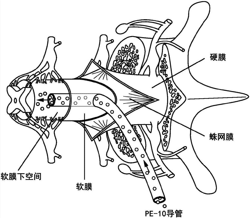
Spinal subpial gene delivery system
A gene delivery, pial technology, applied in gene therapy, genetic engineering, muscular system diseases, etc., can solve problems such as invasiveness
- Summary
- Abstract
- Description
- Claims
- Application Information
AI Technical Summary
Problems solved by technology
Method used
Image

Examples
Embodiment 1
[0089] Materials and methods
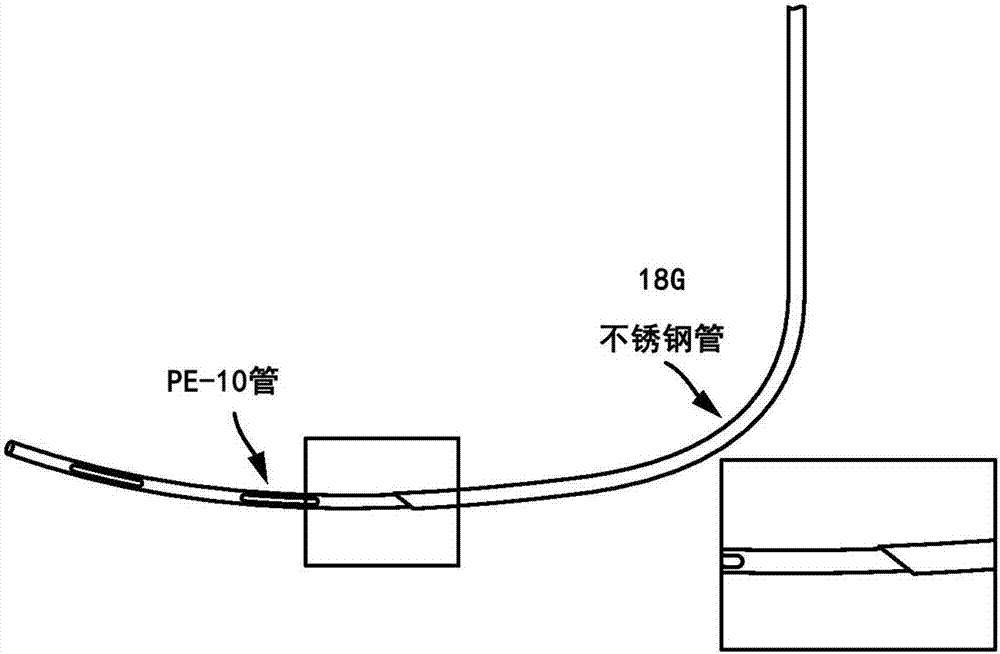
[0090] Animals and general surgical preparation - use adult Sprague-Dawley rats (male and female, 250-350 g; n = 16) or cross-bred from Minnesota and Göttingen strains Adult minipigs (both sexes; 30-40 kg; n=6). Rats were anesthetized with 5% isoflurane and maintained at 2-3% isoflurane during surgery depending on respiration rate and paw pinch response. Then, the hair on the rat's back was shaved and washed with 2% chlorhexidine. Following skin incision, the paraspinal muscles surrounding the cervical, thoracic, or lumbar spine were removed, and the animal was secured into a spinal cord fixation stand (Stoelting) using a Cunningham spinal cord clamp as previously described (Kakinohana et al. People (2004). Region-specific cell grafting into cervical and lumbar spinal cord in rat: aqualitative and quantitative stereological study. Experimental neurology 190: 122-132). To expose the spinal cord, a dorsal laminectomy of the corresponding vertebr...
Embodiment 2
[0098] Substantial AAV9-mediated transgene expression following a single subpial bolus
[0099] Initially, the efficacy of a single bolus subpial delivery of AAV9-UBI-GFP or AAV9-UBI-RFP was tested in rats and pigs. Animals received 20 μl (rats) or 200 μl (pig) AAV9 vector. At 6-8 weeks after AAV9 delivery, spinal cords were dissected from 4% paraformaldehyde-fixed animals and imaged in situ using the Avis fluorescence system. Transverse or horizontal spinal cord sections were then dissected from AAV9 injected segments and analyzed for the presence of GFP or RFP and co-stained with neuronal (NeuN) and glial (GFAP) antibodies. In rat and porcine spinal cords, strong GFP or RFP expression was found on the surface of the spinal cord and was easily identified by visual inspection as yellow-green or red areas. Figure 1H and 1J The picture shows the same as the blank control spinal cord ( Figure 1I ) compared to that in the transected porcine spinal cord and in the anterior r...
Embodiment 3
[0104] GFP expression in distal spinal cord segments
[0105] Next, the descending spinal tract GFP in the lumbar spinal cord was characterized following subpial injection of AAV9-UBI-GFP into the subpial space of the midthoracic (Th6-7) or lower cervical segments in rats and pigs degree of expression. Strong GFP expression was found throughout the entire lumbar spinal cord at 3-6 weeks after subpial AAV9 delivery. Using transverse lumbar (L2-L6) spinal cord sections taken from pigs, high-intensity GFP expression in transected axons in the lateral and ventral cords was easily identified without additional GFP immunostaining ( Figure 4A , a white asterisk). In these regions, similar densities of GFP+ axons were found throughout the white matter. Relatively lower numbers of GFP+ axons were found in the dorsal cord compared to the lateral and ventral cord ( Figure 4A , DF). Consistent with the extent of axonal labeling found in the white matter of the lateral and ventral c...
PUM
 Abstract
Description
Claims
Application Information
 - Generate Ideas
- Intellectual Property
- Life Sciences
- Materials
- Tech Scout
- Unparalleled Data Quality
- Higher Quality Content
- 60% Fewer Hallucinations
Browse by: Latest US Patents, China's latest patents, Technical Efficacy Thesaurus, Application Domain, Technology Topic, Popular Technical Reports.
© 2025 PatSnap. All rights reserved.Legal|Privacy policy|Modern Slavery Act Transparency Statement|Sitemap|About US| Contact US: help@patsnap.com