Medical ultrasonic image segmentation method with high precision and combining global and local regional information

An ultrasound image and local area technology, applied in the field of medical image processing, can solve the problems of strict initial position requirements, boundary leakage, noise and clutter sensitivity, etc., and achieve the effect of strong capture ability, strong noise immunity, and good segmentation effect.

Inactive Publication Date: 2017-06-06
LIAONING NORMAL UNIVERSITY
View PDF8 Cites 3 Cited by
  • Summary
  • Abstract
  • Description
  • Claims
  • Application Information

AI Technical Summary

Problems solved by technology

The active contour model based on global area information has poor ability to capture detailed information, and usually cannot handle heterogeneous objects well, and the evolution speed of the model is slow; the model based on local information can handle the above problems well, However, the model is sensitive to noise and clutter, prone to boundary leakage and has strict requirements on the initial position

Method used


Image

  • Medical ultrasonic image segmentation method with high precision and combining global and local regional information
  • Medical ultrasonic image segmentation method with high precision and combining global and local regional information
  • Medical ultrasonic image segmentation method with high precision and combining global and local regional information

Examples


Embodiment Construction

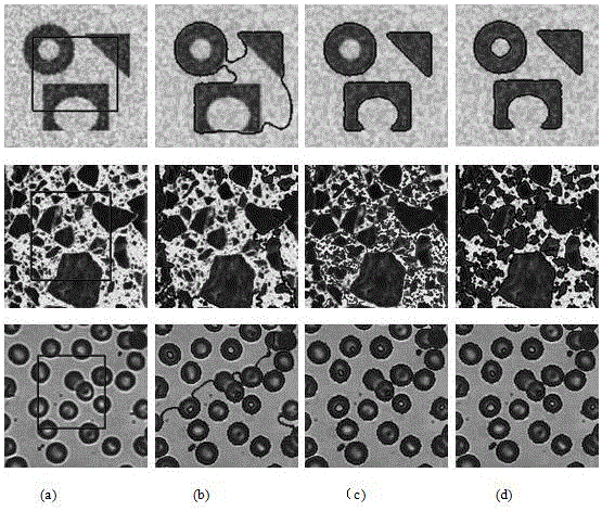
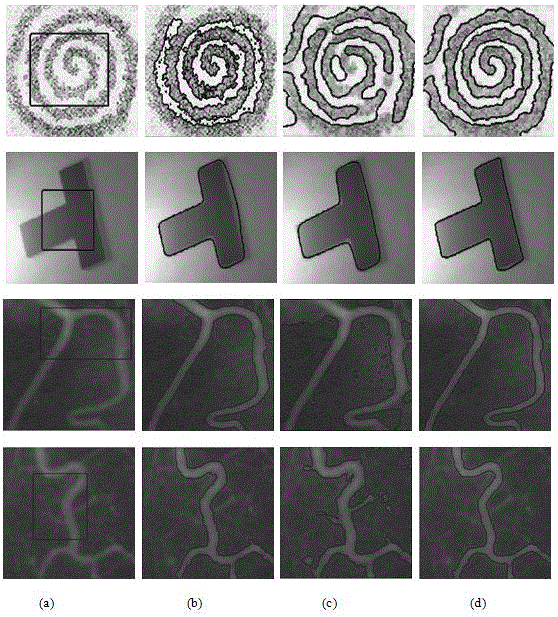
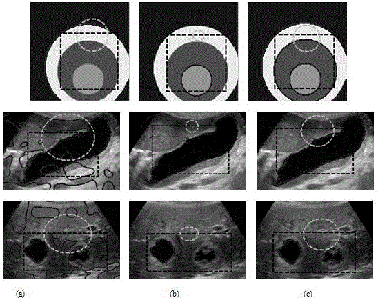
[0029] The medical ultrasonic image segmentation method of the present invention in combination with global and local area information is carried out according to the following steps:

[0030] Step 1: Create an Ultrasound Image The corresponding mathematical model is as follows:

[0031] (1)

[0032] in, and Describe the noise, weak edge region and heterogeneous region of the ultrasound image respectively, here, and Represents global and local regions The internal and external average gray value of , the corresponding expression is:

[0033] and

[0034] (2)

[0035] in, for ultrasound images your region; is the level set function of function;

[0036] Step 2: In order to find The optimal value of , build the model:

[0037] (3)

[0038] in

[0039] (4)

[0040] is an infinitesimal positive number close to 0;

[0041] Step 3. From the Euler-Lagrange equation, the level set evolution equation of model (4) can be obtained:

[004...


PUM


Abstract

The invention discloses a medical ultrasonic image segmentation method with high precision and combining global and local regional information. The method combines image global information and local information. The global information provides a model with good image noise immunity and a good image edge capturing ability. The local information makes more accurate heterogeneous region segmentation. The combination of the global information and the local information can process an image with complex background and internal structure, and can be used for medical ultrasound image segmentation. An experimental result shows that the method can segment the medical ultrasonic image with low contrast and complex structure in a short time, and can obtain a good segmentation effect for the weak images and the images containing noise.

Description

technical field [0001] The invention relates to the field of medical image processing, in particular to a medical ultrasonic image segmentation method combining global and local area information with high segmentation accuracy. Background technique [0002] Due to the influence of external factors such as medical imaging equipment, uneven illumination, and ray energy scattering, medical ultrasound images generally have the following structural features: (1) gray level heterogeneity in the segmented area; (2) images often only contain white, gray and dark gray target area, while the black area is the background area; (3) Due to the complexity of the internal structure of the human body and the irregularity of cartilage tissue, the edges of the ultrasound image are blurred and the internal structure is complex. However, in the detection of clinical medical treatment, the doctor needs to accurately outline the position and shape of the lesion in the ultrasound image, which brin...

Claims


Application Information

Patent Timeline
IPC IPC(8): G06T7/00G06T7/11
CPCG06T7/0012G06T2207/10132
Inventor 方玲玲王相海
Owner LIAONING NORMAL UNIVERSITY
Why Eureka
Try Eureka