
Level set retinal vessel image segmentation method with shape prior being fused

A retinal blood vessel and image segmentation technology, applied in image analysis, image enhancement, image data processing, etc., can solve problems such as oversensitivity, small blood vessels are easy to break, and blood vessels are too wide

Inactive Publication Date: 2016-11-09
JIANGXI UNIV OF SCI & TECH
View PDF2 Cites 26 Cited by
  • Summary
  • Abstract
  • Description
  • Claims
  • Application Information

AI Technical Summary

Problems solved by technology

[0004] The purpose of the present invention is to address the shortcomings of the existing retinal vessel segmentation methods and provide a level-set retinal vessel image segmentation method with fusion shape prior. Fractures, insufficient segmentation of blood vessel intersections, over-sensitivity to image noise, and crossing of target and background gray values

Method used


Image

  • Level set retinal vessel image segmentation method with shape prior being fused
  • Level set retinal vessel image segmentation method with shape prior being fused
  • Level set retinal vessel image segmentation method with shape prior being fused

Examples


Embodiment Construction

[0062] The present invention will be further described below in combination with specific embodiments.

[0063] Explanation of the experiment: the data of the embodiment involved in the application of the present invention comes from the retinal image of the 12th normal person (12_h) in the HRF database.

[0064] This embodiment includes three steps: retinal vessel image preprocessing, vessel image rough segmentation and vessel image fine segmentation.

[0065] The specific description is as follows:

[0066] 1. Retinal blood vessel image preprocessing

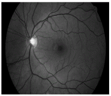
[0067] (a) Select the green channel image I of the retinal image, and use the Geodesic active contours (GAC) model to automatically obtain the "mask" of the retina, such as figure 2 shown.

[0068] (b) Using the retinal "mask" information obtained in the previous step (a), the figure 1 Do edge expansion based on mirror symmetry, and the size of the edge expansion is equal to the size of the Gaussian template in the next s...


PUM


Abstract

The invention relates to a level set retinal vessel image segmentation method with shape prior being fused. The method comprises the following steps: 1) enhancing a retinal vessel image by utilizing a morphological operator and through Gaussian convolution; 2) roughly segmenting the retinal vessel image through anisotropic property of a Hessian matrix and an improved vessel response function, and serving the images as shape constrains and initial information; and 3) constructing a retinal vessel segmentation level set model comprising a local area energy fitting item, a shape constraint item, a level set function regularity maintenance item, a length punishment item and a weight area restraint item by utilizing shape prior and retinal image data. The segmentation method is high in segmentation result accuracy, can replace manual segmentation, and can play an important helping role in diagnosis and treatment of clinical related eye diseases, and has a higher clinical application value.

Description

technical field [0001] The invention relates to a retinal blood vessel image segmentation method based on a level set model, which solves the problems in the existing model that adjacent blood vessels are easily connected, blood vessels are too wide, small blood vessels are easy to break, and blood vessel intersections are insufficiently segmented. Background technique [0002] The retina is an extension of the brain's nervous tissue, with a complex multi-layered organizational structure, and its vascular lesions are one of the important causes of blindness. The level set method is a powerful tool to solve the problem of curve evolution, and its topological adaptability is strong. It can provide a fast and high-accuracy extraction method of retinal blood vessels, and provide help for clinical ophthalmologists in the diagnosis and treatment of diseases. In the field of ophthalmology, information such as the number, branch, angle, and width of retinal blood vessels can be use...

Claims


Application Information

Patent Timeline
IPC IPC(8): G06T7/00G06T5/00
CPCG06T2207/30041G06T2207/20192G06T5/77
Inventor 梁礼明黄朝林陈新建曾璐周发助
Owner JIANGXI UNIV OF SCI & TECH
Learn More