Image Segmentation Method, Pulmonary Nodule Detection Method and Computer Aided Detection System

A nodule and medical image technology, applied in the field of medical image processing, can solve the problems of pulmonary nodules that cannot be identified, cannot accurately identify pulmonary nodules, and inaccurate segmentation results, so as to facilitate diagnosis and analysis and reduce calculation Quantity and versatility

Active Publication Date: 2019-06-28
SHANGHAI UNITED IMAGING HEALTHCARE
View PDF6 Cites 0 Cited by
  • Summary
  • Abstract
  • Description
  • Claims
  • Application Information

AI Technical Summary

Problems solved by technology

[0003] In the prior art, the level set algorithm or the multi-scale threshold method are used for nodule segmentation, but because pulmonary nodules have various forms such as solid nodules, mixed ground-glass nodules, and ground-glass nodules, it is impossible to simply pass the morphological Method to accurately identify pulmonary nodules, especially mixed ...

Method used


Image

  • Image Segmentation Method, Pulmonary Nodule Detection Method and Computer Aided Detection System
  • Image Segmentation Method, Pulmonary Nodule Detection Method and Computer Aided Detection System
  • Image Segmentation Method, Pulmonary Nodule Detection Method and Computer Aided Detection System

Examples


Embodiment Construction

[0047] In the following description, numerous specific details are set forth in order to provide a thorough understanding of the present invention. However, the present invention can be implemented in many other ways different from those described here, and those skilled in the art can make similar extensions without violating the connotation of the present invention, so the present invention is not limited by the specific implementations disclosed below.

[0048] The present invention is described in detail by means of schematic diagrams. When describing the embodiments of the present invention in detail, for convenience of description, the schematic diagrams are only examples, which should not limit the protection scope of the present invention.

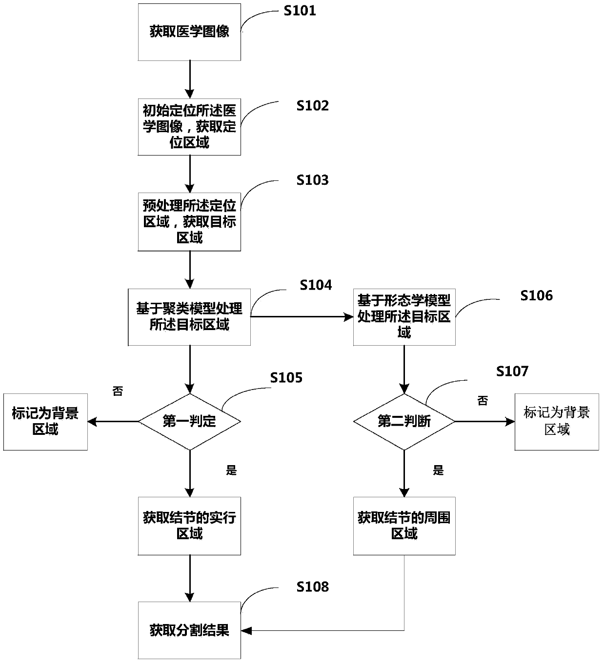
[0049] In order to solve the technical problem of effectively and accurately segmenting different types of pulmonary nodules in the prior art, and improve the accuracy of the user's diagnosis and analysis of the lesion, an image seg...


PUM


Abstract

The invention relates to an image segmentation method, a pulmonary nodule detection method and a computer-aided detection system. The image segmentation method comprises the following steps: a lung CT image is acquired; initial positioning is carried out on the CT image to obtain one or more positioning areas; a filter is adopted to enhance the positioning areas to acquire a target area; the target area processed based on a clustering model is judged, if a first judgment is met, a real region for the nodule is acquired; the target area processed based on a morphological model is judged, if a second judgment is met, a surrounding region for the nodule is acquired; and the real region and the surrounding region are fused to acquire an image segmentation result. According to the method and the system provided by the invention, different types of nodules can be accurately segmented, and follow-up diagnostic analysis can be effectively improved.

Description

【Technical field】 [0001] The invention relates to the field of medical image processing, in particular to an image segmentation method, a pulmonary nodule detection method and a computer-aided detection system thereof. 【Background technique】 [0002] Lung cancer is the cancer with the highest mortality rate worldwide. Although the level of medical diagnosis and treatment continues to improve, the five-year survival rate of lung cancer is only about 15%. Early detection and early treatment are the main ways to improve the cure rate of lung cancer. Pulmonary nodules are an early manifestation of lung cancer. CT scanning is currently the most effective imaging method for lung cancer screening. With the application of Computed Tomography (LDCT), the detection rate of small pulmonary nodules and early lung cancer has been continuously improved. The key technology to realize the automatic analysis and recognition of pulmonary nodules is: the research and application of a series...

Claims


Application Information

Patent Timeline
IPC IPC(8): G06T7/00G06T7/12G06T7/136
CPCG06T7/0012G06T2207/10081G06T2207/30064
Inventor 王季勇
Owner SHANGHAI UNITED IMAGING HEALTHCARE
Why Eureka
Try Eureka