
Adhesion hemocyte image segmentation method based on improved fractional differential and graph theory

A technology of fractional differentiation and image segmentation, which is applied in image analysis, image enhancement, image data processing, etc., can solve the problems of multiple redundant areas, the inability to effectively suppress the generation of small areas, and the difficulty of man-made control of the k value. The effect of broad application prospects

Inactive Publication Date: 2016-07-27
FUZHOU UNIV
View PDF5 Cites 7 Cited by
  • Summary
  • Abstract
  • Description
  • Claims
  • Application Information

AI Technical Summary

Problems solved by technology

However, it also has its own disadvantages. The preset k value in the algorithm is difficult to be effectively controlled artificially. If the value is too large, over-merging will occur; if it is too small, it will not be able to effectively suppress the generation of small areas, and more redundant area

Method used


Image

  • Adhesion hemocyte image segmentation method based on improved fractional differential and graph theory
  • Adhesion hemocyte image segmentation method based on improved fractional differential and graph theory
  • Adhesion hemocyte image segmentation method based on improved fractional differential and graph theory

Examples


Embodiment Construction

[0037] The present invention will be further described below in conjunction with the accompanying drawings and embodiments.

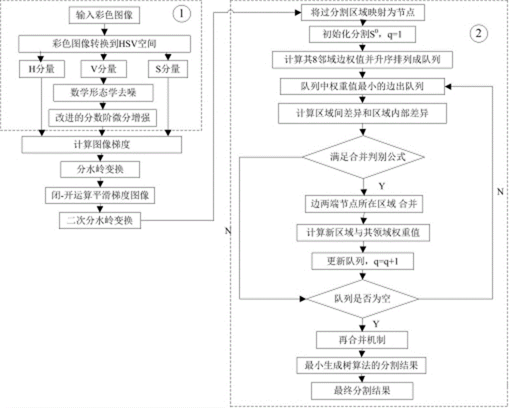
[0038] Such as figure 1 As shown, this embodiment provides a method for segmenting cohesive blood cell images based on improved fractional differential and graph theory, including the following steps:

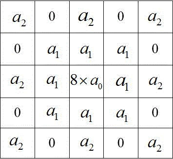
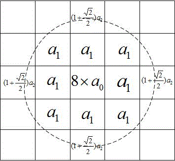
[0039] Step S1: Aiming at the blurred and low-contrast phenomenon of the blood cell image, the blood cell image is preprocessed by combining the morphological denoising and the improved fractional order differential algorithm of the circle-like mask operator. The improved fractional order differential algorithm The algorithm better preserves the details of the cell edge while filtering out the staining pollution and particle noise of the blood cell image;

[0040] Step S2: Use the watershed algorithm to initially segment the preprocessed image, and map the over-segmented regions into nodes;

[0041] Step S3: The cell image obtained in step S2 is re-seg...


PUM


Abstract

The invention relates to an adhesion hemocyte image segmentation method based on the improved fractional differential and graph theory.The method comprises the steps of pretreating a hemocyte image by combining morphological denoising with the improved quasi-circular mask operator fractional differential algorithm aiming at the phenomenon that the hemocyte image is fuzzy and low in contrast ratio, wherein by the adoption of the improved fractional differential algorithm, cell edge details are reserved while staining contamination and grain noise of the hemocyte image are filtered out; then conducting preliminary segmentation on the pretreated image with the watershed algorithm, and mapping an over-segmentation area into a node; finally conducting resegmentation on the cell image obtained in the second step with the improved graph theory minimum spanning tree (MST) algorithm.By the adoption of the method, segmentation precision of adherent cells in the cell image can be improved.

Description

technical field [0001] The invention relates to the technical field of medical image segmentation, in particular to an image segmentation method of cohesive blood cells based on improved fractional differential and graph theory. Background technique [0002] Cells are the basic building blocks of all living organisms. An important direction of biomedicine, which has developed rapidly in recent years, is to diagnose diseases by identifying and counting cells and whether the texture image is distorted. Whether the cell image data can be analyzed accurately depends on whether the cell image can be segmented accurately. [0003] The segmentation method based on graph theory has been a research hotspot at home and abroad in recent years. In 2006, Sharon et al. proposed a top-down hierarchical segmentation method based on graph method in "Nature". The segmentation results are accurate and efficient. Vanhamel et al. proposed a nonlinear multi-scale color image segmentation algori...

Claims


Application Information

Patent Timeline
Patent Type & Authority Applications(China)
IPC IPC(8): G06T7/00G06T5/00
CPCG06T2207/20192G06T2207/20152G06T5/70
Inventor 林丽群王卫星
Owner FUZHOU UNIV
Learn More