Medical ultrasonic esophagoscope tube, and medical ultrasonic esophagoscope
A technology of ultrasound and esophagoscopy, applied in the fields of ultrasound/sonic/infrasonic Permian technology, ultrasound/sonic/infrasonic image/data processing, organ movement/change detection, etc., which can solve the problem of tight fit and inability to form sound transmission Windows, poor image quality, etc.
- Summary
- Abstract
- Description
- Claims
- Application Information
AI Technical Summary
Problems solved by technology
Method used
Image


Examples
Embodiment 1
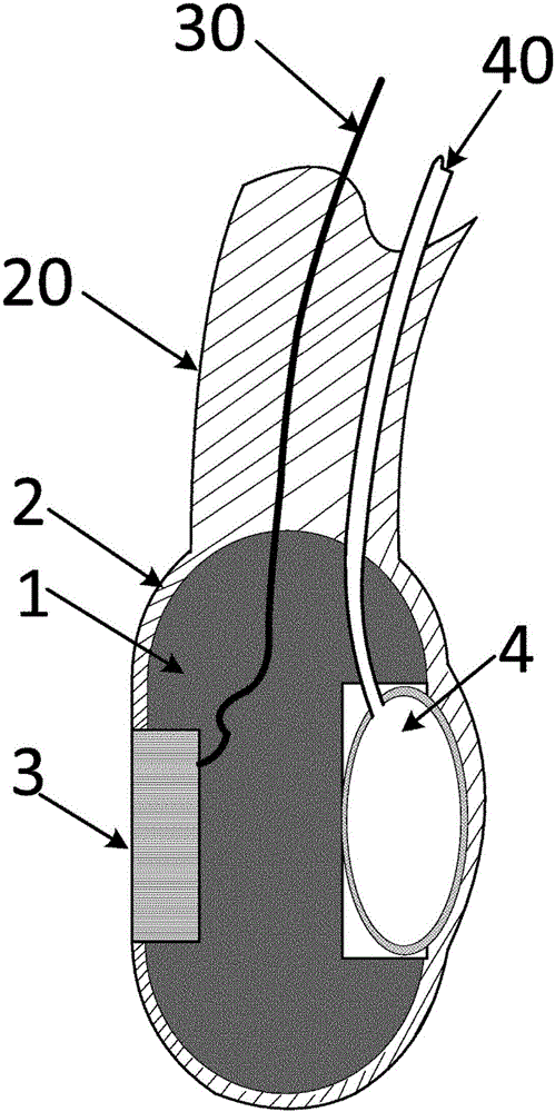
[0106] Implementation example 1, such as Figure 1-2 As shown, a medical ultrasonic esophagoscope mirror tube is characterized in that it includes a head frame 1, a head cuff 2, an ultrasonic probe 3, a radial air bag 4, an elastic extension 20, and is used for the control and information of the ultrasonic probe 3. Electrical channels 30 for exchange, fluid channels 40 for radial airbag 4 control;
[0107] The head skeleton 1 is a hard entity;
[0108] The head cuff 2 is an elastic membrane;
[0109] The ultrasonic probe 3 is fixedly installed in the head frame 1, and the working surface of the ultrasonic probe 3 faces outward;
[0110] The head cuff 2 covers the outer surface of the head frame 1, and the head cuff 2 does not cover the working surface of the ultrasonic probe 3;
[0111] The elastic extension part 20 is fixedly connected with the head frame 1 and the head cuff 2, and the elastic extension part 20 has good elasticity;
[0112] The electrical channel 30 for t...
Embodiment 2
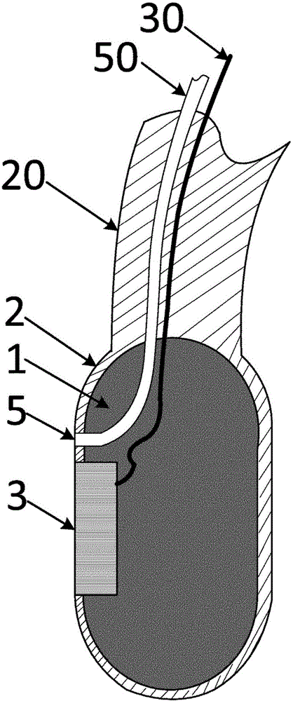
[0115] Implementation example 2, such as image 3 As shown, a medical ultrasonic esophagoscope mirror tube is characterized in that it includes a head frame 1, a head cuff 2, an ultrasonic probe 3, a radial air bag 4, an elastic extension 20, and is used for the control and information of the ultrasonic probe 3. The electrical channel 30 for exchange, the medium hole 5, the medium channel 50 for the input medium of the medium hole 5;
[0116] The head skeleton 1 is a hard entity;
[0117] The head cuff 2 is an elastic membrane;
[0118] The ultrasonic probe 3 is fixedly installed in the head frame 1, and the working surface of the ultrasonic probe 3 faces outward;
[0119] The head cuff 2 covers the outer surface of the head frame 1, and the head cuff 2 does not cover the working surface of the ultrasonic probe 3;
[0120] The elastic extension part 20 is fixedly connected with the head frame 1 and the head cuff 2, and the elastic extension part 20 has good elasticity;
[...
Embodiment 3
[0124] Implementation example 3, such as Figure 4 As shown, a medical ultrasonic esophagoscope mirror tube is characterized in that it includes a head frame 1, a head cuff 2, an ultrasonic probe 3, a radial air bag 4, an elastic extension 20, and is used for the control and information of the ultrasonic probe 3. Electrical channel 30 for exchange, fluid channel 40 for radial airbag 4 control, medium hole 5, medium channel 50 for medium hole 5 input medium;
[0125] The head skeleton 1 is a hard entity;
[0126] The head cuff 2 is an elastic membrane;
[0127] The ultrasonic probe 3 is fixedly installed in the head frame 1, and the working surface of the ultrasonic probe 3 faces outward;
[0128] The head cuff 2 covers the outer surface of the head frame 1, and the head cuff 2 does not cover the working surface of the ultrasonic probe 3;
[0129] The elastic extension part 20 is fixedly connected with the head frame 1 and the head cuff 2, and the elastic extension part 20 h...
PUM
 Abstract
Description
Claims
Application Information
 - R&D
- Intellectual Property
- Life Sciences
- Materials
- Tech Scout
- Unparalleled Data Quality
- Higher Quality Content
- 60% Fewer Hallucinations
Browse by: Latest US Patents, China's latest patents, Technical Efficacy Thesaurus, Application Domain, Technology Topic, Popular Technical Reports.
© 2025 PatSnap. All rights reserved.Legal|Privacy policy|Modern Slavery Act Transparency Statement|Sitemap|About US| Contact US: help@patsnap.com