
Method for constructing individualized human urethra tissue digital model

A technology of digital model and urethra, applied in the medical field, can solve problems such as low imaging effect, low resolution of soft tissue structure, and inability to achieve full coverage of patients

Inactive Publication Date: 2015-11-11
SHANGHAI SIXTH PEOPLES HOSPITAL
View PDF0 Cites 0 Cited by
  • Summary
  • Abstract
  • Description
  • Claims
  • Application Information

AI Technical Summary

Problems solved by technology

Although enhanced CT has many advantages such as ultra-high resolution and thin-slice imaging, it is limited by its low resolution of soft tissue structure. Effective acquisition of image information of cavernous tissue
Although MRI technology can have a better display effect on soft tissues, its imaging effect is slightly lower than that of MDCT.
More importantly, due to the characteristics of MRI, its scope of application is limited to a certain extent, and it is impossible to cover all patients

Method used


Image

  • Method for constructing individualized human urethra tissue digital model
  • Method for constructing individualized human urethra tissue digital model

Examples


Embodiment 1

[0039] In step 1.1, the patient to be tested is placed in a supine position, and the penis-scrotum area and the external urethral orifice area are disinfected.

[0040]Stretch the penis in a weak state, and inject 10ml of tetracaine mucilage from the outer opening of the urethra to block the outer opening of the urethra and prevent the glue from overflowing.

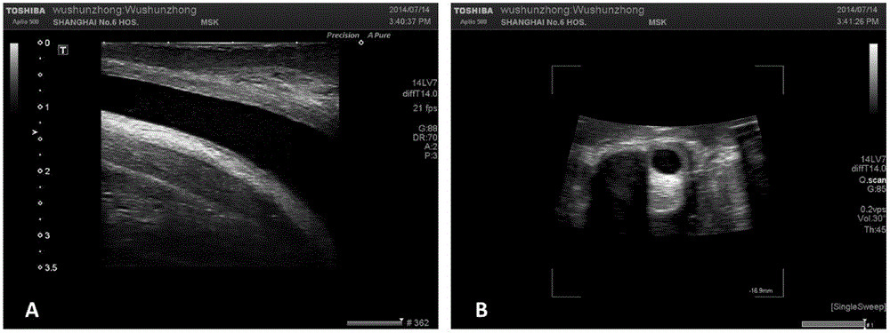
[0041] Step 1.2, using Toshiba Aplio500 ultrasonic diagnostic instrument, probe model PLT-1204MV (center frequency 14MHz). Place the ultrasound probe at the penis-scrotum junction to observe the condition of the urethral lumen.

[0042] If the ultrasound finds that the urethral lumen is not fully opened, or the opening is not satisfactory. You can continue to inject tetracaine glue until the inner diameter of the urethra is greater than 8mm.

[0043] Such as figure 1 As shown in , the dynamic scanning mode is used to scan the urethra and its surrounding tissues to obtain coronal urethral image information. Data is sa...


PUM


Abstract

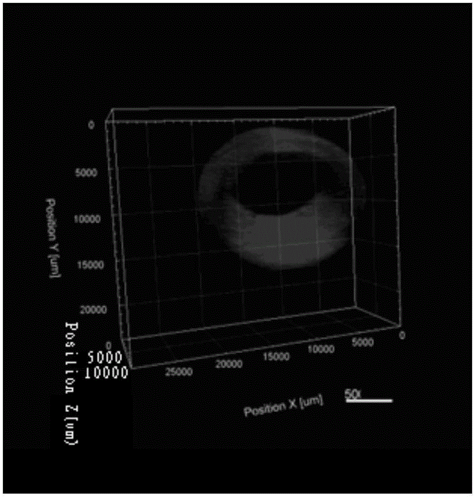
The invention discloses a method for constructing an individualized human urethra tissue digital model. The method comprises the following steps: thin-layer penis overall imaging information of a patient is obtained through a three-dimensional ultrasound technology; an image area to be constructed in the overall imaging information is screened by a manual outline drawing method; and the image area to be constructed undergoes three-dimensional reconstruction and processing by a three-dimension technology, and an individualized urethra three-dimensional digital model for 360-degree observation is constructed. The model constructed by the technical scheme fits the physiological anatomy position structure of normal human urethra, contains all information required by a three-dimensional model and accords with patients' personal characteristics.

Description

technical field [0001] The invention belongs to the field of medical technology, and relates to a method for constructing a digital model of human tissue in medicine, in particular to a method for constructing an individualized digital model of human urethral tissue by using three-dimensional ultrasonic imaging tools. Background technique [0002] In the past ten years, the attention paid to the treatment of urethral stricture in complicated cases is showing a trend of continuous heating up. Although the current clinical gold standard for urethral repair and replacement surgery is still to use various autologous tissues such as penile flap, buccal mucosa, tongue mucosa and even colonic mucosa for reconstruction. However, the introduction of the concept of tissue engineering in this field has caused more and more impact on the status of this gold standard. A series of successful reports of basic and clinical research at home and abroad show that it may become the gold standa...

Claims


Application Information

Patent Timeline
Patent Type & Authority Applications(China)
IPC IPC(8): G06T17/00
Inventor 陈磊冯超李喆吕向国胡兵徐月敏
Owner SHANGHAI SIXTH PEOPLES HOSPITAL
Learn More