Enhanced Vascular Visualization Using a Robotically Steered Endoscope

A speculum and blood vessel technology, applied in the field of visualization systems, can solve problems such as slowing down separation

Active Publication Date: 2017-01-18
KONINKLJIJKE PHILIPS NV
View PDF8 Cites 0 Cited by
  • Summary
  • Abstract
  • Description
  • Claims
  • Application Information

AI Technical Summary

Problems solved by technology

Since the LIMA is removed from the mid-lower portion of the chest, the LIMA tends to become more embedded in the tissue, slowing dissection and making visualization of arteries and collaterals more challenging

Method used


Image

  • Enhanced Vascular Visualization Using a Robotically Steered Endoscope
  • Enhanced Vascular Visualization Using a Robotically Steered Endoscope
  • Enhanced Vascular Visualization Using a Robotically Steered Endoscope

Examples


Embodiment Construction

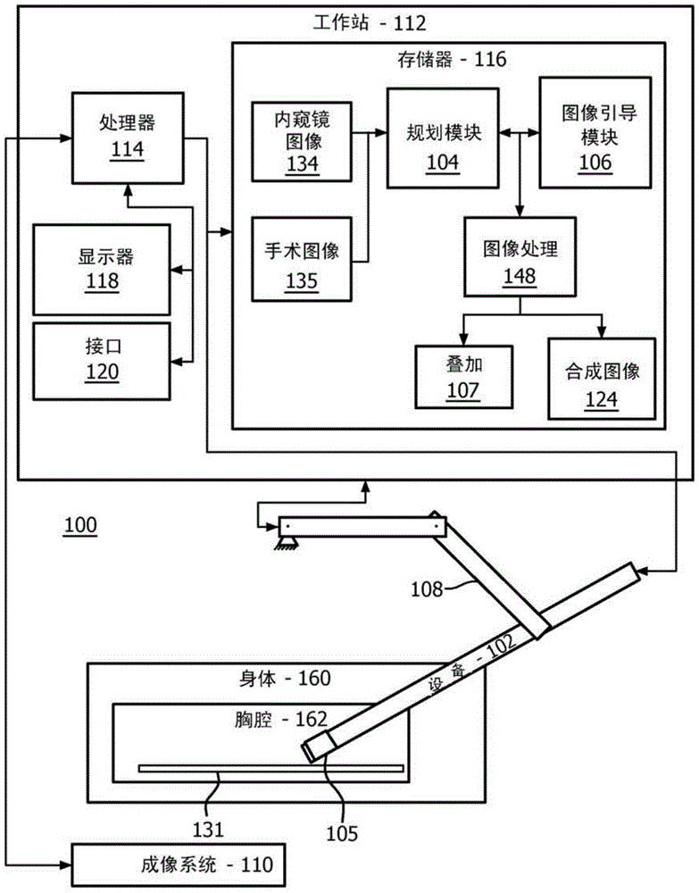
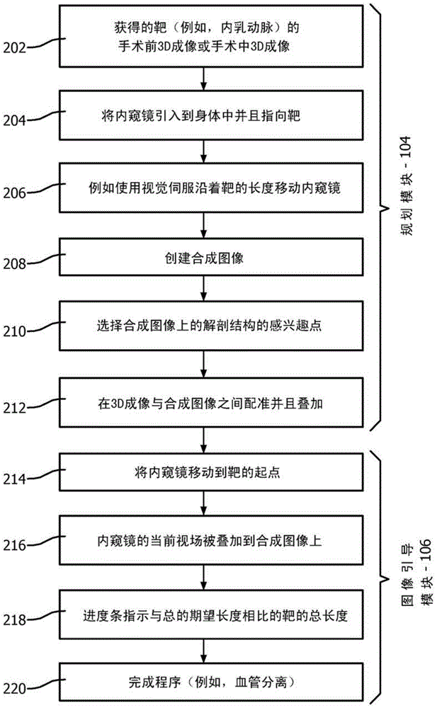
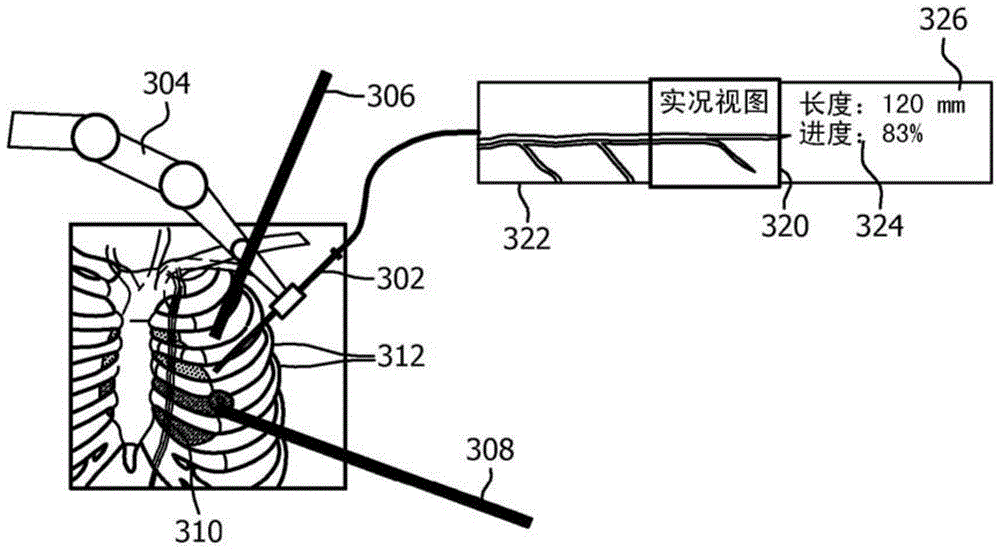
[0024] According to the present principle, a blood vessel separation planning and operation system is provided to solve the above-mentioned problem of blood vessel separation. The present principles provide a significantly enlarged field of view showing a large portion of a vessel (eg LIMA) and provide additional information about the vessel as opposed to only showing a small segment. This additional information includes, for example, the length of the vessel to be separated for harvesting, the progress of vessel separation with respect to the desired bypass length, and the location of side branches that need to be cauterized.

[0025] It should be understood that the present invention will be described in terms of a medical instrument for use with and for a coronary artery bypass procedure; however, the teachings of the present invention are much broader and are applicable where a target anatomy is required or desired Enhanced visualization of any instrument or program. In s...


PUM


Abstract

A system for visualizing target anatomy comprising: a speculum (102) for internal imaging having a field of view smaller than a region to be imaged; a planning module (104) configured to a scope receiving video such that the field of view images of the scope are stitched together to generate a composite image of the region to be imaged; an image guidance module (016) configured to follow the target during the procedure The anatomy moves the scope such that images generated in the field of view of the scope are displayed as live video superimposed on the field of view of the composite image.

Description

technical field [0001] The present disclosure relates to medical instruments and methods, and more particularly to systems and methods for improved visualization of internal anatomy in medical applications. Background technique [0002] Coronary artery bypass grafting (CABG) is a surgical procedure used to revascularize blocked coronary arteries. In a conventional surgical procedure, the patient's sternum is opened and the heart is fully exposed. However, minimally invasive (MI) bypass surgery is performed through small incisions. An important part of the CABG procedure is the removal of blood vessels from the patient's body, which are then used to bypass one or more atherosclerotic narrowings in the coronary arteries. The most commonly removed and used vessel is the internal mammary artery (IMA), which may include the left internal mammary artery (LIMA) or the right internal mammary artery (RIMA) located in the chest cavity. [0003] Direct access to these vessels is not...

Claims


Application Information

Patent Timeline
Patent Type & Authority Patents(China)
IPC IPC(8): A61B1/00A61B1/313
CPCA61B1/00006A61B1/00009A61B1/00045A61B1/00147A61B1/00149A61B1/00172A61B1/005A61B1/04A61B1/3132A61B5/489A61F2/062G06T11/60G06T2210/41A61B2034/302A61B34/30A61B2090/365A61B1/000094
Inventor A·波波维奇H·埃尔哈瓦林
Owner KONINKLJIJKE PHILIPS NV
Why Eureka
Try Eureka