
Fluorescence in situ hybridization method for metaphase chromosomes of mulberry

A fluorescence in situ hybridization and chromosome technology, which is applied in the field of genetics, can solve the problems such as the application of fluorescence in situ hybridization technology, chromosome expansion and deformation, and chromosome production failure, etc., and achieves clear signal points, good shape and stable number. Effect

Active Publication Date: 2016-01-06
SOUTHWEST UNIV
View PDF2 Cites 0 Cited by
  • Summary
  • Abstract
  • Description
  • Claims
  • Application Information

AI Technical Summary

Problems solved by technology

However, in the process of FISH chromosome preparation, the pretreatment, the concentration of probes and antibodies, the temperature and time of co-denaturation of probes and chromosomes all affect the experimental results. For example, whether the pretreatment is appropriate is the most critical operation in chromosome preparation technology. The steps determine whether the chromosome length is moderate and whether it is conducive to hybridization; if hypotonicity and insufficient or excessive enzymatic hydrolysis will lead to the failure of chromosome preparation; and if the concentration of probes and antibodies is too high, it will easily lead to incomplete elution and non-specificity False positive results from hybridization; the temperature and time of co-denaturation of probes and chromosomes are also very critical. If the co-denaturation temperature is low or the time is short, the double strands are not fully opened, and no specific signal will be hybridized, and the co-denaturation temperature is too high or the time is too high Too long will cause chromosome expansion and deformation, which is not conducive to obtaining normal experimental results
FISH technology has been successfully used in many plants, but so far there has been no report on the application of fluorescence in situ hybridization technology in mulberry

Method used


Image

  • Fluorescence in situ hybridization method for metaphase chromosomes of mulberry
  • Fluorescence in situ hybridization method for metaphase chromosomes of mulberry
  • Fluorescence in situ hybridization method for metaphase chromosomes of mulberry

Examples


Embodiment 1

[0026] The fluorescence in situ hybridization method of mulberry metaphase chromosome comprises the following steps:

[0027] (1) Preparation of chromosomal slide specimens: using the young leaves of Chuanmulus mulberry as materials, the chromosomal slide specimens were prepared by the wall-removing hypotonic flame-drying method. The specific steps are as follows:

[0028] The young leaves of Chuan mulberry were pretreated in 8-hydroxyquinoline aqueous solution with a concentration of 0.002M at 25°C in the dark for 3 hours, and then fixed at 4°C for more than 2 hours with methanol: glacial acetic acid volume ratio of 3:1. Rinse 3 times with distilled water again; Then the fixed mulberry leaves are hypotonic at 25° C. for 30 minutes in 1 / 15M aqueous KCl solution, rinsed 2 times with distilled water and then soaked with 5% (W / V) cellulase and 5% (W / V) Enzymolysis at 25°C for 2 hours in a mixed solution of (W / V) pectinase, then rinsed with distilled water for 3 times and stayed i...

Embodiment 2

[0039] The fluorescence in situ hybridization method of mulberry metaphase chromosome comprises the following steps:

[0040] (1) the preparation of chromosome slide specimen, the preparation method is identical with embodiment 1;

[0041] (2) 25SrDNA probe preparation: according to the conserved region sequence of 25SrDNAgenes, design primers for amplifying 25SrDNA in the conserved region, as follows:

[0042] 25SrDNA forward primer: 5'-ccaaatgcctcgtcatctaa-3' (SEQ ID NO.3);

[0043] 25SrDNA reverse primer: 5'-gcgaatcaacggttcctct-3' (SEQ ID NO.4);

[0044] Then use the Chuanmulus genomic DNA as a template, and use SEQIDNO.3 and SEQIDNO.4 as primers to carry out PCR amplification to obtain the nucleotide sequence shown in SEQIDNO.6, and then connect it with pMD19-Tsimplevector to obtain pMD19-25SrDNA;

[0045] The obtained recombinant plasmid pMD19-25SrDNA is used as a template, and the PCRDIGProbeSynthesisKit (Roche) kit is used to prepare 25SrDNA probes. The amplification ...

Embodiment 3

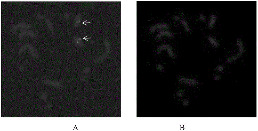
[0048] Example 3 is the same as Example 1 for the method of fluorescence in situ hybridization of mulberry metaphase chromosomes, except that the probe added in the hybridization solution is a mixed probe of a biotin-labeled 5SrDNA probe and a digoxin-labeled 25SrDNA probe The preparation method of the biotin-labeled 5SrDNA probe is the same as the preparation method of the 5SrDNA probe in Example 1, the difference is that the PCR BiotinProbeSynthesisKit (Roche) is used to prepare the probe, which contains Biotin-dUTP. Anti-digoxigenin-fluorescein (Anti-digoxigenin-fluorescein, Fabfragments (from sheep)) with a concentration of 2 ng / μL from sheep was used to incubate for 1 hour, and then labeled with Cy3 at a concentration of 10 ng / μL. anti-biotin antibody (Cy TM 3-Streptavidin Conjugate (ZyMAX TM Grade)) was incubated for 1 h, and the results of microscopic examination were as follows: image 3 shown.


PUM


Abstract

The invention discloses a fluorescence in situ hybridization method for metaphase chromosome of mulberry. The fluorescence in situ hybridization method comprises the following steps: the enzymolysis wall degaradation hypotonic flame drying method is adopted to prepare a chromosome slide sample, so that a high-quality mulberry chromosome flake can be obtained; then 5S rDNA and 25S rDNA probes are prepared, and the probes are in repetitive sequence, strong in fluorescence signal, easy to detect and high in repeatability; finally, fluorescence in situ hybridization of the chromosome is conducted to obtain the mulberry chromosome in complete form. The fluorescence in situ hybridization method can be used for mulberry chromosome identification and structure research, and provides a new way for evolution research and the like of mulberry.

Description

technical field [0001] The invention belongs to the field of genetics, and in particular relates to a fluorescence in situ hybridization method for metaphase chromosomes of mulberry trees. Background technique [0002] Mulberry (Morusalba L.) is a plant of the genus Moraceae. As an important economic tree, its leaves are the main feed for silkworms. Mulberry trees have been cultivated for more than 7,000 years. However, most common mulberry trees are cultivated species propagated by cuttings and grafting. The genetic background is relatively vague, which makes mulberry trees encounter great obstacles in the study of genetic breeding and evolutionary analysis. At present, conventional karyotype analysis of mulberry chromosomes has been reported, and mulberry trees have rich types of chromosome ploidy changes, for example, Sichuan mulberry (M.notabilis) has 14 chromosomes, Indian mulberry (M.indica), white mulberry (M.alba) There are 28 chromosomes in M. bombycis, 42 in M. bo...

Claims


Application Information

Patent Timeline
Patent Type & Authority Patents(China)
IPC IPC(8): C12Q1/68
CPCC12Q1/6841C12Q2563/131
Inventor 何宁佳李杨徐云敏向仲怀
Owner SOUTHWEST UNIV
Learn More