Method and magnetic resonance device for the automated classification of an image property of a magnetic resonance image

A technology of magnetic resonance imaging and magnetic resonance equipment, applied in the direction of using nuclear magnetic resonance imaging system for measurement, magnetic resonance measurement, and magnetic variable measurement

Active Publication Date: 2014-05-21
SIEMENS HEALTHCARE GMBH
View PDF6 Cites 3 Cited by
  • Summary
  • Abstract
  • Description
  • Claims
  • Application Information

AI Technical Summary

Problems solved by technology

The coil can only tr...

Method used


Image

  • Method and magnetic resonance device for the automated classification of an image property of a magnetic resonance image
  • Method and magnetic resonance device for the automated classification of an image property of a magnetic resonance image
  • Method and magnetic resonance device for the automated classification of an image property of a magnetic resonance image

Examples


Embodiment Construction

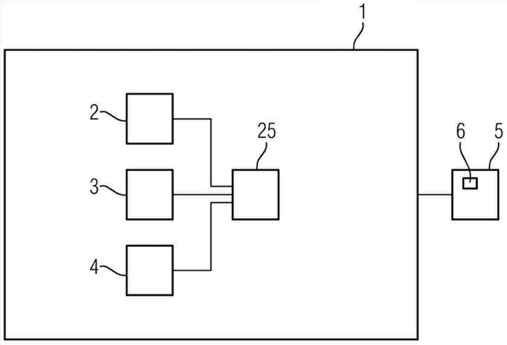
[0053] figure 1 A magnetic resonance system 1 according to the invention is shown schematically. The magnetic resonance system is designed as a magnetic resonance tomography system, ie in particular has the coils required for generating the magnetic field gradient. The magnetic resonance system includes a simulation unit 2 , a classification unit 3 and a storage unit 4 . A display unit 5 is provided for displaying the magnetic resonance image 6 . Furthermore, elements obviously present in the magnetic resonance tomography system, such as coils for generating the main magnetic field B0, the patient table, control and circuit electronics, etc., are not shown in detail and are assumed to be known and available.

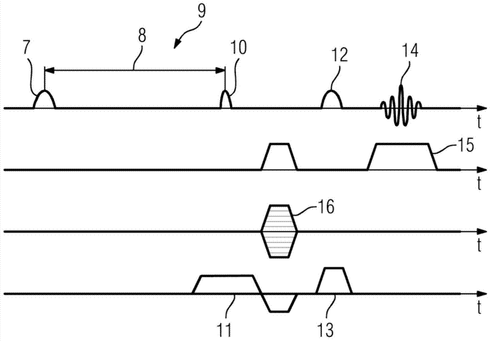
[0054] figure 2 Sequence diagram showing the FLAIR image capture sequence. The HF pulse sequence and the magnetic field gradient used to capture k-space lines are represented as follows:

[0055] First, the 180° inversion pulse 7 is incident. This is followed by a...


PUM


Abstract

A method is disclosed for the automated classification of an image property of a magnetic resonance image(6). In an embodiment, the method includes determining at least the parameters T1, T2 and proton density of at least two tissues imaged in the magnetic resonance image(6) or prespecifying at least two sets of at least these parameters; calculating the signal intensity in the center of the k-space for each imaged tissue or each set of parameters; and classifying the image property as a function of the calculated signal intensities. A magnetic resonance device is also disclosed.

Description

technical field [0001] The invention relates to a method and a magnetic resonance system for automatic classification of image features of magnetic resonance images. Background technique [0002] Since the invention of rapid imaging, magnetic resonance imaging has evolved into a standard method in medical diagnosis. There are various recording methods which differ, inter alia, with respect to the required measurement time and the resulting image contrast. There are methods for recording so-called spin-density or proton-density-weighted, T1-, T2- or T2*-weighted images, and diffusion imaging is also known. [0003] When the following refers to the recording of images, this clearly means that, as usual in magnetic resonance tomography, the slices located in the object under examination of the magnetic resonance tomography system are excited with the aid of excitation pulses and slice-selective gradients, the detection coils are used to The signal of the excited spins is read...

Claims


Application Information

Patent Timeline
IPC IPC(8): G01R33/56G01R33/563A61B5/055
CPCG01R33/5608G01R33/50G01R33/5602
Inventor T.费威尔
Owner SIEMENS HEALTHCARE GMBH
Why Eureka
Try Eureka