
Method for neuroepithelial cells differentiation from pluripotent stem cells and medium using same

A technology for neuroepithelial cells and induction medium, applied in cell culture active agents, biochemical equipment and methods, embryonic cells, etc. The effect of shortening the differentiation time

Active Publication Date: 2013-06-19
鸿雁生技有限公司
View PDF2 Cites 7 Cited by
  • Summary
  • Abstract
  • Description
  • Claims
  • Application Information

AI Technical Summary

Problems solved by technology

Another scholar added two smad inhibitors (smad inhibitors) (Elkabetz et al., 2008; Lee et al., 2007; Chambers et al., 2009) to the stem cell differentiation process, including Noggin and SB431542, to shorten neural differentiation time; some scholars also use genetic manipulation to induce differentiation or co-culture with other cells to induce differentiation (Pankratz et al., 2007; Chambers et al., 2009). However, although the above methods can obtain neuroepithelial cells, they still cannot make large Some embryonic stem cells differentiate into nerve cells, and have disadvantages such as expensive production costs, safety concerns about using viruses for genetic manipulation, and uncertain factors in co-cultivation with other cells
In addition, if the neuroepithelial cells produced are mixed with other non-neural cells or undifferentiated cells, it may affect the subsequent neural differentiation, and the undifferentiated pluripotent stem cells may also become teratomas when transplanted into animals

Method used


Image

  • Method for neuroepithelial cells differentiation from pluripotent stem cells and medium using same
  • Method for neuroepithelial cells differentiation from pluripotent stem cells and medium using same
  • Method for neuroepithelial cells differentiation from pluripotent stem cells and medium using same

Examples


Embodiment 1

[0056] Example 1: Making embryonic stem cells into embryoid bodies

[0057] Human embryonic stem cells TW1 were cultured at 37°C, 5% CO 2 Then the cultured embryonic stem cells were cultured as small cell aggregates in suspension culture in DMEM-F12 medium containing 20% ​​serum substitute (knock-out replacement serum; KSR; Invitrogen, USA), and 37°C, 5% CO 2 The conditions were cultured for 2 days, so that the embryonic stem cells coalesced into cell clusters of embryoid bodies (embryoid bodies).

Embodiment 2

[0058] Example 2: Neuroepithelial cell induction differentiation phase

[0059] The cell mass of the embryoid body in Example 1 was transferred to a 15 ml centrifuge tube, and the supernatant was aspirated after allowing the cells to settle at room temperature. Prepare 500 mL of the first neural induction medium, the composition of which is shown in Table 1 below, and add the drug Bio at a concentration of 0.5 μM (0.5 μmol / L, the same below), the drug SB431542 at a concentration of 10 μM, and the drug SB431542 at a concentration of 10 ng / mL Fibroblast growth factor 2; It should be noted that the individual concentrations of the components of the first neural induction medium are not limited to those described above, specifically, the concentration of the drug Bio can range from 0.05 μM to Between 50 μM, the concentration range of the drug SB431542 is between 1 μM and 100 μM, and the concentration range of the fibroblast growth factor 2 is between 1 ng / mL and 100 ng / mL.

[006...

Embodiment 3

[0063] Example 3: Neuroepithelial cell induction differentiation phase

[0064] Take the neuroepithelial cells in Example 2, replace the first neural induction medium with the second neural induction medium, and add fibroblast growth factor 2 with a concentration of 10 ng / mL, and update the second neural induction medium every 2 days. The second nerve induction medium is used to further differentiate the neuroepithelial cells, wherein the composition of the second nerve induction medium is shown in Table 2 below.

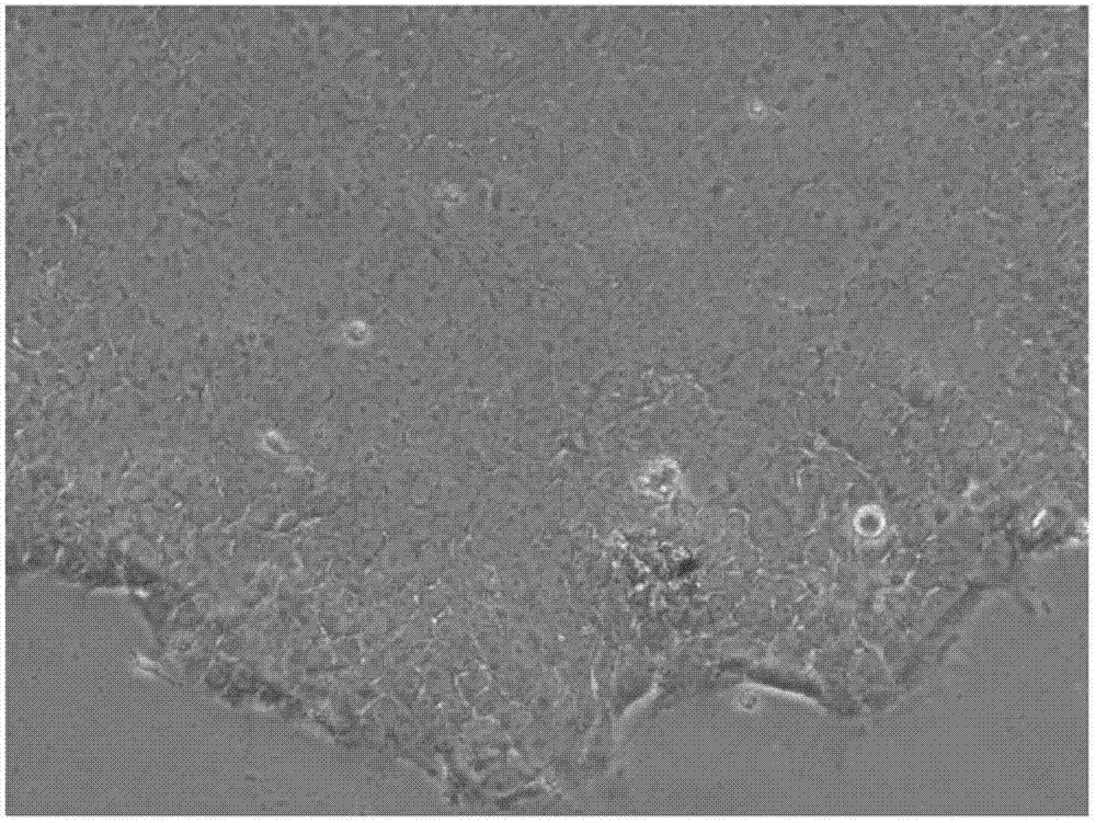
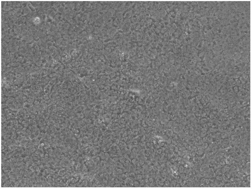
[0065] The cultured and differentiated cells were observed under a microscope with a microscope magnification of 100, 200 and 400 times, and the results are shown in Figure 3 and Figure 4, wherein, by Figure 3A It can be seen that the uniform shape of the cells and the tight columnar shape around the spherical structure; Figure 3B It can be seen that the cells have a spherical structure, and the surroundings are in a tight cylindrical shape and the central part i...


PUM

PropertyMeasurementUnit
concentrationaaaaaaaaaa

Abstract

The present invention discloses a neural induction medium comprising Wnt-signal agonist, TGFbeta-signal inhibitor and FGF-signal agonist for inducing neural differentiation. The neural induction medium used in a culture system is capable for inducing the neural differentiation of stem cells into neuroepithelial cells which are useful for the clinical applications. Therein, the neuroepithelial cells can further differentiate into mature neurons for the practical applications including regeneration medicine and drug discovery for neural disorders.

Description

technical field [0001] The present invention relates to a method for differentiating neuroepithelial cells and the medium used therefor, in particular to a method for differentiating neuroepithelial cells differentiated from pluripotent stem cells and the medium used therefor. Background technique [0002] Stem cells refer to primitive cells that have not yet fully differentiated, but have the ability to self-renew and can differentiate into two or more mature cells. Stem cells are classified according to their differentiation ability, and can be divided into totipotent stem cells, pluripotent stem cells, multipotent stem cells and bipotent stem cells; Can be divided into embryonic stem cells (embryonic stem cells), adult stem cells (somatic stem cells) and induced pluripotent stem cells (induced pluripotent stem cells; iPSC). Among them, human embryonic stem cells are pluripotent stem cells, which come from the inner cell mass of the blastocyst stage before implantation. T...

Claims


Application Information

Patent Timeline
Patent Type & Authority Applications(China)
IPC IPC(8): C12N5/079
CPCC12N5/0623C12N2501/115C12N2501/415C12N2506/02
Inventor 苏鸿麟陈圣美
Owner 鸿雁生技有限公司
Learn More