Human residual ear cartilage stem cells and method for constructing tissue engineered cartilage
A technique for chondrocyte and tissue engineering, applied in the fields of medicine and biomedical engineering
- Summary
- Abstract
- Description
- Claims
- Application Information
AI Technical Summary
Problems solved by technology
Method used
Image
Examples
preparation example Construction
[0063] The preparation method of the tissue-engineered cartilage of the present invention is simple and convenient. A certain amount of the human remnant ear chondrocytes is mixed with a pharmaceutically acceptable biodegradable material, and then the cartilage is induced in vitro. The method comprises the steps of:
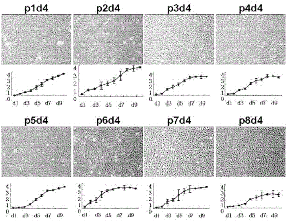
[0064] In the first step, the cells derived from human residual ear cartilage are cultured and amplified, and subcultured to 1-8 generations;
[0065] In the second step, the above-mentioned subcultured and expanded cells are centrifuged to form pellets or high-density seeding to form cell membranes; or these cells are mixed with solid medically acceptable biodegradable materials to form cell-biomaterial composites thing;
[0066] In the third step, the agglomerates, membrane sheets or complexes are placed in chondrogenic induction medium for in vitro culture.
[0067] The method of planar culture expansion in the first step is, for example but not limited to, ...
Embodiment 1
[0103] Acquisition and culture of cartilage stem cells from human remnant ear
[0104] The cells are taken from the autologous residual ear cartilage tissue of patients with microtia. The donors are 8-30 years old, healthy, and free from malignant tumors, infectious diseases and autoimmune diseases.
[0105] (1) Material collection: Under aseptic conditions in the operating room, after satisfactory anesthesia, drape was routinely sterilized, and the patient's residual ear tissue was cut out, placed in a 50mL sterile centrifuge tube, soaked in 40mL sterile saline for storage and transport.
[0106] (2) Separation of residual ear chondrocytes: Under sterile conditions in an ultra-clean bench, completely remove the connective tissue and perichondrium around the residual ear cartilage, and chop the cartilage to 1-2mm 3 Small pieces were soaked in 0.25% chloramphenicol solution for 30-40min, rinsed with PBS, digested with 0.25% trypsin + 0.02% EDTA (Amresco, USA) in a constant temp...
Embodiment 2
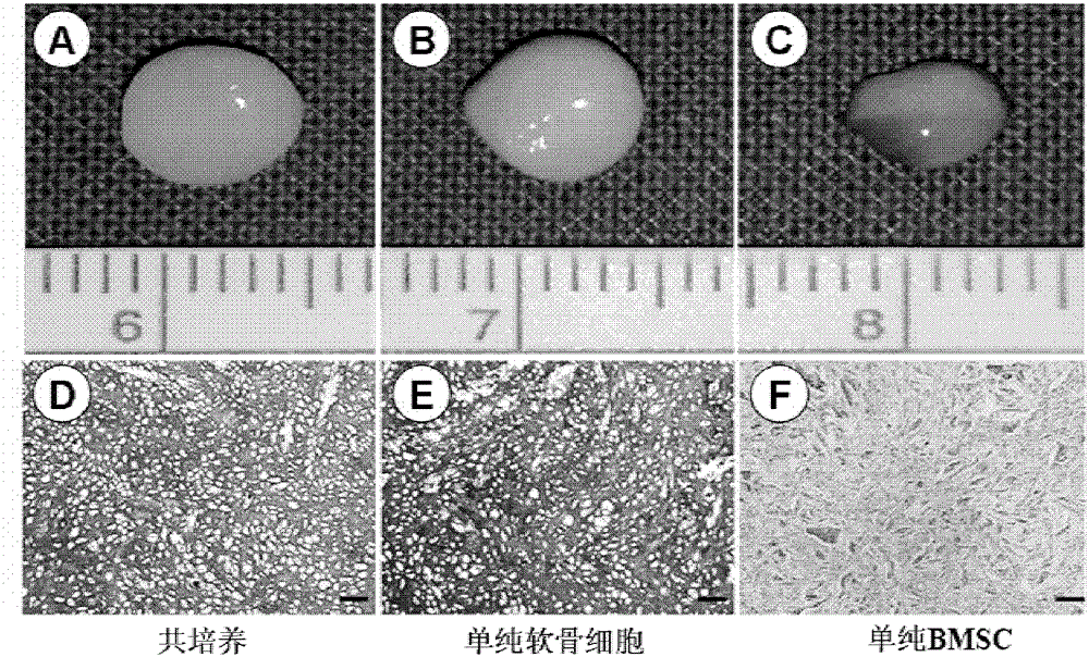
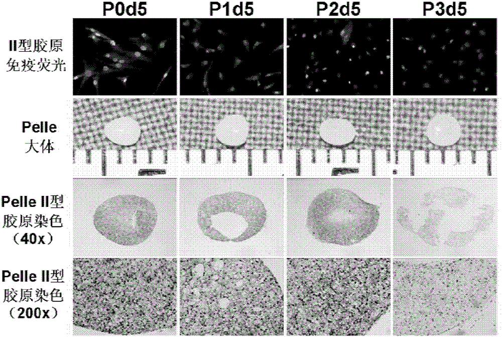
[0112] Preparation of pellets from remnant ear cartilage stem cells
[0113] (1) Cell collection: when the monolayer cultured human residual ear chondrocytes reached 90% confluence, they were digested with 0.25% trypsin+0.02% EDTA. After the cells were collected, they were counted with a cell counter. Trypan blue staining showed that the number of viable cells was more than 90%. The cells were centrifuged at 1800 rpm for 8 min to precipitate the cells. The supernatant was discarded, and the cells were dispersed by shaking.
[0114] (2) Preparation of pellets: After collecting the cells, resuspend them in ordinary culture medium to prepare 0.4×10 pellets. 6 / ml of cell suspension. Take 1ml of the suspension in a 15ml centrifuge tube and centrifuge at 1000rpm for 5min to make a cell pellet. Slightly loosen the bottle cap and place at 37°C, 5% CO 2 , cultured in an incubator with saturated humidity, and then changed the medium every other day (ordinary culture medium or chondr...
PUM
 Abstract
Description
Claims
Application Information
 - R&D
- Intellectual Property
- Life Sciences
- Materials
- Tech Scout
- Unparalleled Data Quality
- Higher Quality Content
- 60% Fewer Hallucinations
Browse by: Latest US Patents, China's latest patents, Technical Efficacy Thesaurus, Application Domain, Technology Topic, Popular Technical Reports.
© 2025 PatSnap. All rights reserved.Legal|Privacy policy|Modern Slavery Act Transparency Statement|Sitemap|About US| Contact US: help@patsnap.com


