
Aortic valve ultrasonic image segmentation method based on probability distribution and continuous maximum flow

An aortic valve and ultrasound image technology, applied in the field of image processing, can solve the problems of ultrasound image resolution, low contrast, long time consumption, calcification of valve leaflets and valve annulus, etc.

Active Publication Date: 2013-01-16
SHANGHAI JIAO TONG UNIV
View PDF2 Cites 10 Cited by
  • Summary
  • Abstract
  • Description
  • Claims
  • Application Information

AI Technical Summary

Problems solved by technology

However, due to the following characteristics of the medical ultrasound image of the aortic valve, its segmentation has become a difficult problem: 1) low resolution and low contrast of the ultrasound image; 2) the inherent speckle noise of the ultrasound image; Wave texture characteristics; 4) The movement of the three leaflets caused by the opening and closing of the aortic valve; 5) Artifacts caused by severe calcification of the leaflets and annulus
The threshold segmentation method is simple in principle and easy to operate, but due to the inherent speckle noise and texture characteristics in the above-mentioned ultrasound images, the segmentation results are very poor, and it is difficult to provide accurate and effective guidance information for clinical treatment
Based on the doctor's manual segmentation method, because the doctor has long-term clinical practice experience, the segmented results can be further used in clinical diagnosis and treatment, but because of the heavy workload and long time-consuming, it does not have good application value

Method used


Image

  • Aortic valve ultrasonic image segmentation method based on probability distribution and continuous maximum flow
  • Aortic valve ultrasonic image segmentation method based on probability distribution and continuous maximum flow
  • Aortic valve ultrasonic image segmentation method based on probability distribution and continuous maximum flow

Examples


Embodiment

[0043] This embodiment is implemented in a computer with a CPU of Pentuim IV 2.6GHz, a graphics card of NVIDIA Geforce GTX 460, a memory of 2.0GB, and a programming language of C++.

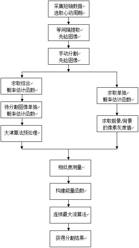
[0044] The implementation process of this embodiment is as follows figure 1 shown.

[0045] The first step is to collect medical ultrasound image data of the short axis of the human aortic valve, select a continuous and complete cardiac cycle, and extract five frames of prior images at equal intervals. At this time, each frame of prior image will represent a difference in a cardiac cycle. Phase;

[0046] The second step is to manually segment the above five frames of prior images, and calculate the bounding box of each frame segmentation result, and take the largest bounding box for the subsequent process;

[0047] The third step is to calculate a comprehensive center point of the prior image according to the respective independent center points of the prior image segmentation results, with the g...


PUM


Abstract

The invention relates to an aortic valve ultrasonic image segmentation method based on probability distribution and continuous maximum flow. The method comprises the following steps: 1, acquiring medical ultrasonic image data of a human body aortic valve short axis, and extracting a five-frame prior image at equal intervals; 2, segmenting the five-frame prior image; 3, constructing a two-dimensional gray-distance histogram; 4, calculating to obtain a comprehensive probability estimation function through the two-dimensional gray-distance histogram; 5, respectively calculating a respective independent probability estimation function; 6, respectively calculating the pixel gray values which can respectively represent the foreground and background for the five-frame prior image; 7, solving an independent probability estimation map for the current image to be segmented; 8, respectively measuring the similarity for the foreground area and the manual segmentation result of the five-frame prior image; and 9, obtaining the segmentation result. Compared with the prior art, the aortic valve ultrasonic image segmentation method is stable, reliable, convenient to implement and suitable for actual clinical application.

Description

technical field [0001] The invention relates to the technical field of image processing, in particular to an aortic valve ultrasonic image segmentation method based on probability distribution and continuous maximum flow. Background technique [0002] Medical ultrasound imaging has attracted extensive attention due to its unique advantages, such as real-time, non-invasive, repeatable, high sensitivity, and low cost. In the clinical diagnosis and treatment of the aortic valve based on medical ultrasound images, according to the different pathological conditions of the patients, it is necessary to extract the information of the aortic valve, and one of the important methods is image segmentation. The quality of the image segmentation results directly affects the positioning, quantitative and qualitative analysis of lesion tissue structure, 3D reconstruction and other follow-up operations, as well as the accuracy of treatment planning adopted by image-guided surgery and tumor r...

Claims


Application Information

Patent Timeline
IPC IPC(8): G06T7/00A61B8/08
Inventor 顾力栩聂媛媛罗哲
Owner SHANGHAI JIAO TONG UNIV
Learn More