
Separation method of cancer stem cells

A technology of tumor stem cells and separation methods, which is applied in the field of cell separation, can solve the problems of inapplicable separation and limitations of tumor stem cells, and achieve the effect of good application prospects

Active Publication Date: 2011-04-20
THE SECOND AFFILIATED HOSPITAL ARMY MEDICAL UNIV
View PDF0 Cites 7 Cited by
  • Summary
  • Abstract
  • Description
  • Claims
  • Application Information

AI Technical Summary

Problems solved by technology

Current studies have found that in some tumor cell populations, the SP phenotype is not suitable for the isolation of tumor stem cells; moreover, because Hoechst33342 itself has a certain cytotoxic effect, it also limits the application of this method

Method used


Image

  • Separation method of cancer stem cells
  • Separation method of cancer stem cells
  • Separation method of cancer stem cells

Examples


Embodiment Construction

[0025] Hereinafter, preferred embodiments of the present invention will be described in detail with reference to the accompanying drawings.

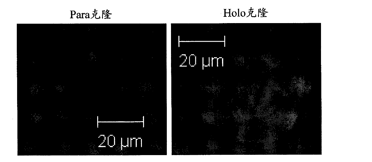
[0026] Based on some existing clues: (1) Mitochondrial membrane potential is closely related to cell differentiation state; (2) Mitochondrial membrane potential is related to differentiation potential and tumorigenicity of normal stem cells; (3) Mitochondrial membrane potential of tumor cells Higher than normal cells; the inventor speculates that: associated with their special biological properties, a subset of cancer stem cells may have a mitochondrial membrane potential different from that of differentiated tumor cells. In a preferred embodiment, the inventors used different methods to find that the mitochondrial membrane potential of lung cancer stem cell subsets was significantly higher than that of differentiated lung cancer cells; after sorting lung cancer cell subsets with different mitochondrial membrane potentials by flow cytomet...


PUM


Abstract

The invention discloses a separation method of cancer stem cells. Cancer stem cells and differentiated tumor cells can be separated by different mitochondrial membrane potential of different cells. The cell subgroup with high mitochondrial membrane potential is rich in cancer stem cells, and the cell subgroup with low mitochondrial membrane potential is rich in differentiated tumor cells.

Description

technical field [0001] The invention relates to a cell separation method, in particular to a tumor stem cell separation method. Background technique [0002] More and more evidences show that the tumor cell population is a heterogeneous cell population, among which only the tumor stem cell subgroup has the ability of unlimited proliferation, self-renewal, multidirectional differentiation and tumorigenesis, which is the key factor for the occurrence, progression and recurrence of malignant tumors. Therefore, it is of great scientific significance to study the special biological characteristics of tumor stem cells in depth. [0003] However, isolation of CSCs from tumor cell populations remains difficult due to their scarcity. At present, there are mainly three methods for the isolation of tumor stem cells: (1) Fluorescence-activated flow cytometry and magnetic cell sorting based on surface markers such as CD133, CD44, and CD34. However, since most of the surface markers com...

Claims


Application Information

Patent Timeline
IPC IPC(8): C12N5/06C12N5/08G01N21/64
Inventor 余时沧钱桂生叶小群王光辉黄桂君
Owner THE SECOND AFFILIATED HOSPITAL ARMY MEDICAL UNIV
Learn More