Skull coronal CT (computed tomography) scanning fixator

A CT scanning, coronal technology, applied in computed tomography scanners, patient positioning for diagnosis, echo tomography, etc. Effect

Inactive Publication Date: 2011-04-20
LONGGANG DISTRICT CENT HOSPITAL OF SHENZHEN
View PDF5 Cites 2 Cited by
  • Summary
  • Abstract
  • Description
  • Claims
  • Application Information

AI Technical Summary

Problems solved by technology

It cannot satisfy the patient’s fixation when the head is lifted up to the state where the mandible is almost perpendicular to the scanning table when the patient is in the prone position.

Method used


Image

  • Skull coronal CT (computed tomography) scanning fixator
  • Skull coronal CT (computed tomography) scanning fixator

Examples


Embodiment Construction

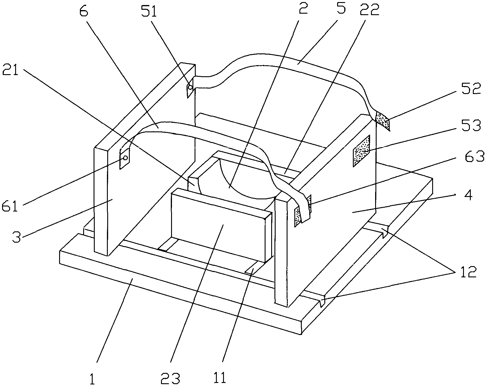
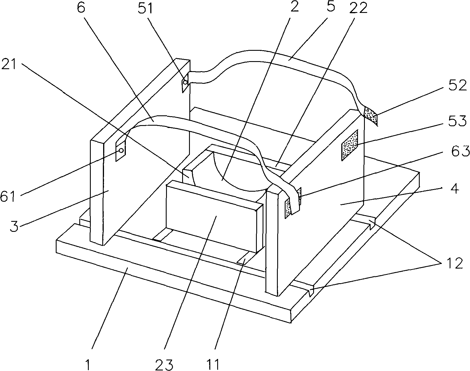
[0026] From the accompanying drawings, it can be clearly seen that the cranial coronal CT scanning fixator includes: a base plate 1 that can be conveniently fixed on the CT workbench, and the base plate 1 is vertically provided with two left plates that can act on the bilateral buccal surfaces of the skull 3 and the right side plate 4, the bottom plate 1 between the left side plate 3 and the right side plate 4 is provided with a front wall 22 and a rear wall 23 formed by the groove body 21 and the front wall 22 and the rear wall 23 that can seal the groove body 21 front and rear ends. The mandibular positioning groove 2, the left side plate 3 and the right side plate 4 front are provided with a forehead limiting elastic band 5 for the patient’s skull forehead limit, the left side plate 3 and the right side plate 4 rear are provided with a for The back pillow limit elastic belt 6 of patient's head back pillow limit.

[0027] The bottom plate 1 between the left side plate 3 and ...


PUM


Abstract

The invention relates to a skull coronal CT (computed tomography) scanning fixator which comprises a baseplate capable of being conveniently fixed at a CT operation table, a left side plate and a right side plate, which can act on bilateral buccal surfaces of a skull, are vertically arranged on the baseplate, and the fixator is characterized in that a lower jaw positioning groove is arranged on the baseplate between the left side plate and the right side plate; a forehead limiting elastic band for limiting the forehead of the skull of a patient is arranged at the front parts of the left side plate and the right side plate; and an occiput limiting elastic band for limiting the occiput of the skull of the patient is arranged at the back parts of the left side plate and the right side plate. The fixator can effectively comfortably assist the patient in lifting the skull upwards till the lower jaw is almost vertical to a scanning bed and fixed, thereby not only reducing the fatigue of keeping the skull raised upwards of the patient under the state without dependence for a long time, but also effectively solving the problem of CT image artifacts caused by involuntary movement of the skull of the patient. By using the CT scanning fixator, the smooth completion of CT scanning can be favored, and clear and accurate CT scanned images can be obtained, thereby having good application and popularization prospects.

Description

technical field [0001] The invention relates to a medical device, in particular to a cranial coronal CT scan fixer suitable for CT scan operations. Background technique [0002] With the rapid development of medical imaging, CT scanning has become a major means of medical examination. Coronal head CT scanning can observe the structures of the eyeball, sinuses, parietal bone, and saddle area in the coronal direction, and combined with transverse images to diagnose diseases It is of great significance to make a definite diagnosis, and as a special form of scanning, it has gradually shown its important position in imaging diagnosis. [0003] However, during coronal head CT scanning, the patient is usually required to lie prone on the CT scanning table, and the patient is required to keep the head lifted until the mandible is nearly perpendicular to the scanning table until the end of the scan. It is difficult for patients to keep their head up for a long time when there is no ...

Claims


Application Information

Patent Timeline
Patent Type & Authority Applications(China)
IPC IPC(8): A61B6/04A61B6/03
Inventor 胡卫东项立范义鄂占森高永波
Owner LONGGANG DISTRICT CENT HOSPITAL OF SHENZHEN
Why Eureka
Try Eureka