
Method for executing three-dimensional cardiac ultrasound virtual endoscopy

A virtual endoscope and heart technology, applied in surgery, catheters, etc., can solve the problems that cannot meet the requirements of accurate diagnosis of complex congenital heart disease, observers are lost, and traumatic critical patients cannot tolerate it.

Inactive Publication Date: 2010-10-06
XIN HUA HOSPITAL AFFILIATED TO SHANGHAI JIAO TONG UNIV SCHOOL OF MEDICINE +2
View PDF0 Cites 13 Cited by
  • Summary
  • Abstract
  • Description
  • Claims
  • Application Information

AI Technical Summary

Problems solved by technology

Although traditional cardiac catheterization can achieve a more accurate diagnosis, its invasiveness often makes it unbearable for critically ill patients
Two-dimensional echocardiography (2DE) is a non-invasive diagnostic technique, but since complex congenital heart disease is a combination of various intracardiac and extracardiac malformations or defects in three-dimensional space, the shape of the heart and its composition The spatial position relationship of the patient often plays an important role in the diagnosis, and the examiner must perform three-dimensional spatial imagination on a series of two-dimensional images to make a three-dimensional diagnosis, which cannot meet the requirements for accurate diagnosis of complex congenital heart disease in three-dimensional space
[0005] However, the heart is a relatively special organ. The heart of the observed object is in an active state. In each cardiac cycle, the heart is constantly moving. When sampling under the gate control of the electrocardiogram and respiratory cycle, due to sampling errors and the uncooperative patient Due to factors such as loss of data sampling and processing distortion in image reconstruction, it brings certain difficulties to virtual heart observation and measurement. In addition, due to the complex structure of the heart, the observer also suffers from disorientation.

Method used


Image

  • Method for executing three-dimensional cardiac ultrasound virtual endoscopy
  • Method for executing three-dimensional cardiac ultrasound virtual endoscopy
  • Method for executing three-dimensional cardiac ultrasound virtual endoscopy

Examples


Embodiment Construction

[0029] The specific implementation of the method for performing three-dimensional cardiac ultrasound virtual endoscopy provided by the present invention will be described in detail below with reference to the accompanying drawings.

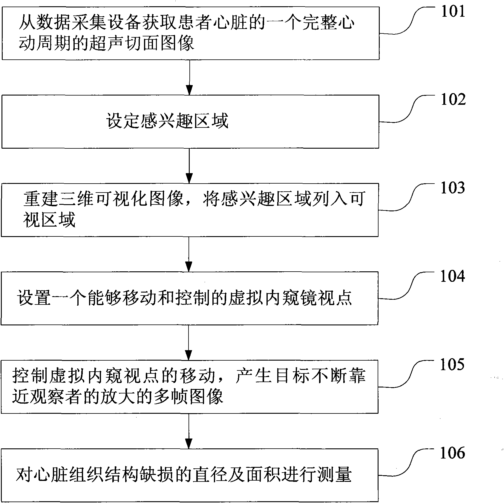
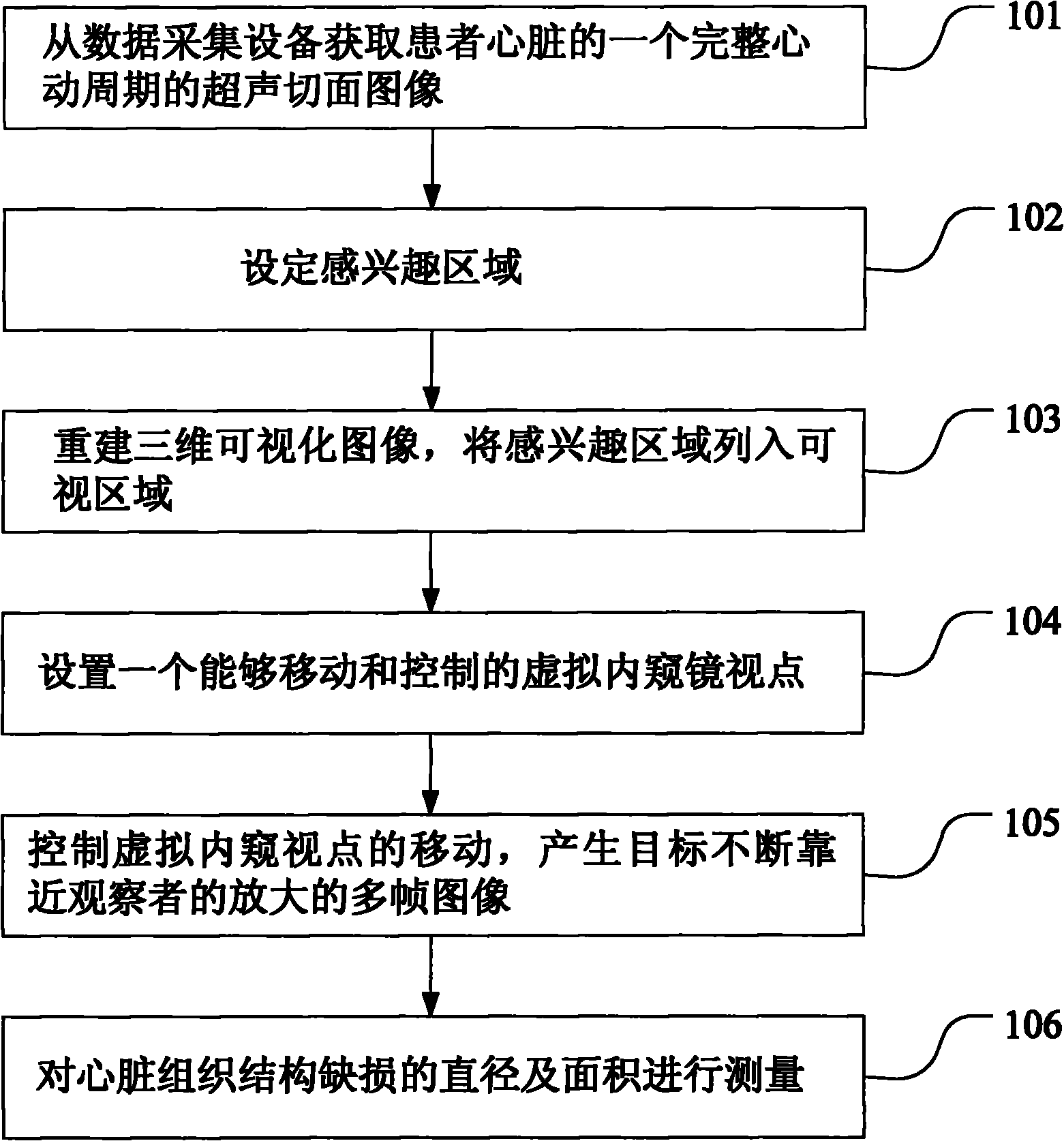
[0030] The specific implementation method is as follows: step S101, obtain an ultrasonic section image of a complete cardiac cycle of the patient's heart from the data acquisition device; step S102, set the region of interest; step S103, reconstruct the three-dimensional visualization image, and include the region of interest into three-dimensional Visualization area; step S104, setting a virtual endoscope viewpoint that can be moved and controlled; step S105, controlling the movement of the virtual endoscope viewpoint, and producing enlarged multi-frame images of the target constantly approaching the observer; The diameter and area of ​​tissue structural defects were measured.

[0031] In step S101, an ultrasonic section image of a complete cardi...


PUM


Abstract

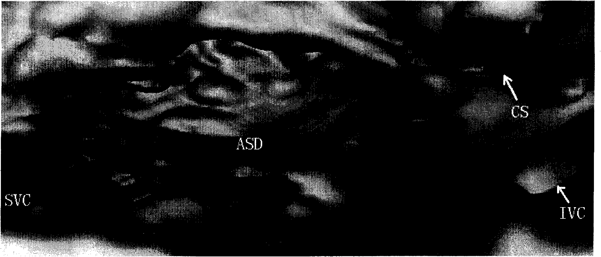
The invention provides a method for executing three-dimensional cardiac ultrasound virtual endoscopy. The method comprises the following steps of: acquiring an ultrasound sectional image on a complete cardiac cycle of a patient heart from data acquisition equipment; acquiring a three-dimensional data set which comprises two-dimensional image information of an overall heat by using the ultrasound sectional image; performing three-dimensional reconstruction according to the data set; setting a moveable and controllable virtual endoscopic viewpoint in a three-dimensional visual image; and controlling the virtual endoscopic viewpoint to move in a three-dimensionally displayed cardiac chamber so as to generate a plurality of frames of magnified images that a structural target in an observed heart is close to an observer continuously in a corresponding cardiac cycle. Due to the adoption of the method, the virtual viewpoint observation position can be converted at any time according to the requirement of the observer; and the observer can observe a movement change situation of a structure in the heart in the complete cardiac cycle.

Description

【Technical field】 [0001] The invention relates to the technical field of computerized medical imaging, in particular to a method for performing three-dimensional cardiac ultrasound virtual endoscopy. 【Background technique】 [0002] Among pediatric diseases, congenital heart disease (CHD) is one of the leading causes of infant death. At least 100,000 to 150,000 children with congenital heart disease are born every year in our country, and complex congenital heart disease accounts for 35-38%, of which 60-80% are in a critical state after birth, because of failure to make accurate anatomical diagnosis in time and early Correction and death. Although traditional cardiac catheterization can achieve a more accurate diagnosis, its invasiveness often makes it unbearable for critically ill patients. Two-dimensional echocardiography (2DE) is a non-invasive diagnostic technique, but since complex congenital heart disease is a combination of various intracardiac and extracardiac malfo...

Claims


Application Information

Patent Timeline
IPC IPC(8): A61B8/12A61B1/00
Inventor 孙锟薛海虹余建国陈滨津王威琪
Owner XIN HUA HOSPITAL AFFILIATED TO SHANGHAI JIAO TONG UNIV SCHOOL OF MEDICINE
Learn More