


Quick Research
Generate reliable direction feasibility study reports for your R&D in just a few steps.

Technical Q&A
Discover and master advanced knowledge NOW. Basics, ideas, possibilities, all at once.

Find Solutions
As an expert in R&D theories, this can generate solutions to your technical problems instantly.

Evaluate Feasibility
Analyze your overall solution with one click, know your potential R&D risks in advance.

Monitor Landscape
Get weekly tech updates, stay abreast of the latest tech innovations and key insights.
Actuator apparatus, image pickup apparatus and endoscopic apparatus
A technology of an actuator device and an imaging device, which is applied in the direction of endoscopes, instruments, telescopes, etc., and can solve the problems of miniaturization of the front end of the endoscope, that is, difficulty in reducing the diameter, difficulty in miniaturization of the connecting part, and poor jointability, etc.
- Summary
- Abstract
- Description
- Claims
- Application Information
AI Technical Summary
Problems solved by technology
Method used
Image


Examples
no. 1 Embodiment approach
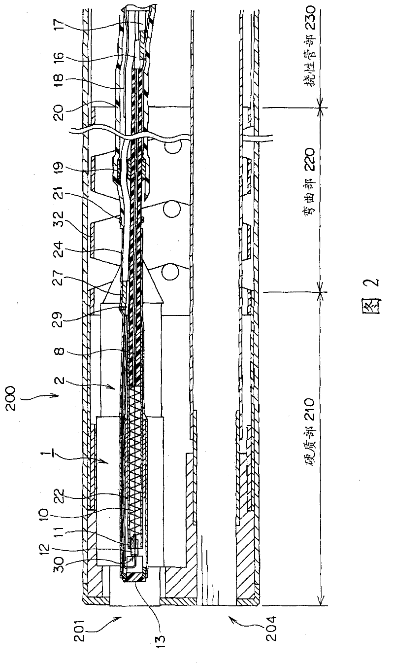
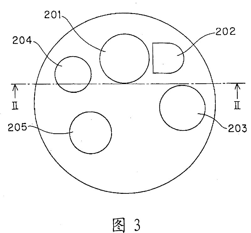
[0053] First, use Figure 1 to Figure 8 The present invention will be described. Figure 1 to Figure 8 Regarding the first embodiment of the present invention, figure 1 It is a structural diagram of the imaging device of the electronic endoscope, figure 2 is along image 3 The profile of the II-II line, image 3 It is the front view of the front end of the endoscope, Figure 4 is along figure 1 Sectional view of line IV-IV, Figure 5 yes Figure 4 Enlarged view of part V of Figure 6 is the structure diagram of the insulating ball, Figure 7 is the structure diagram of other insulating balls, Figure 8 It is an enlarged view of the moving part of the lens.
[0054] In this embodiment, for figure 1 The illustrated imaging unit 1 that moves the internal lens forward and backward will be described. The imaging unit 1 constitutes an imaging device used in an endoscope and is used to realize a focusing function or a zoom / telephoto function. In this imaging unit 1 , an ...
no. 2 Embodiment approach
[0085] Next, use Figure 9 The actuator 2 of the imaging unit 1 of the second embodiment will be described. In addition, in the following description, the same code|symbol is attached|subjected to the same component as 1st Embodiment, and description is abbreviate|omitted, and only a different part is demonstrated. and, Figure 9 The second embodiment of the present invention is equivalent to the first embodiment Figure 4 sectional view.
[0086] In this embodiment, instead of the SMA wire 28 for GND in the first embodiment, a wire 51 made of a conductive material such as stainless steel or steel is used. One end of the wire 51 on the base end side is crimped by the second crimping portion 29 and is electrically connected to the GND cable 27 by soldering. On the other hand, like the SMA wire 8 of the first embodiment, the other end of the front end side of the wire 51 is hooked by the insulating ring 12 and folded back (refer to Figure 6 ), through the insulating pipe 1...
no. 3 Embodiment approach
[0092] Next, use Figure 10 The actuator 2 of the imaging unit 1 of the third embodiment will be described. In addition, in the following description, the same code|symbol is attached|subjected to the same component as 1st and 2nd embodiment, description is abbreviate|omitted, and only a different part is demonstrated. and, Figure 10 The third embodiment of the present invention is equivalent to the first embodiment Figure 4 sectional view.
[0093] The actuator 2 of the present embodiment is configured such that an SMA wire 54 is connected to the GND side of the wire 51 of the second embodiment. In addition, the SMA wire 54 of this embodiment may replace the SMA wire 28 for GND of 1st Embodiment.
[0094] One end of the SMA wire 54 on the proximal side is crimped by the second crimping portion 29 and is electrically connected to the GND cable 27 . In addition, the other end on the front end side of the SMA wire 54 is crimped and connected together with the wire 51 by a...
PUM
 Abstract
Description
Claims
Application Information
 - R&D Engineer
- R&D Manager
- IP Professional
- Industry Leading Data Capabilities
- Powerful AI technology
- Patent DNA Extraction
Browse by: Latest US Patents, China's latest patents, Technical Efficacy Thesaurus, Application Domain, Technology Topic, Popular Technical Reports.
© 2024 PatSnap. All rights reserved.Legal|Privacy policy|Modern Slavery Act Transparency Statement|Sitemap|About US| Contact US: help@patsnap.com