Method for cutting blood-vessel data in digital blood-vessel angiograph image

An angiography and digital subtraction technology, applied in the field of medical imaging, can solve problems such as difficult, unreasonable, and harsh judgments

Inactive Publication Date: 2008-07-23
HUAZHONG UNIV OF SCI & TECH
View PDF4 Cites 0 Cited by
  • Summary
  • Abstract
  • Description
  • Claims
  • Application Information

AI Technical Summary

Problems solved by technology

Since it is not judged whether there are blood vessels in the sub-block, a segmentation result will also be obtained for the background area without blood vessels, which is obviously unreasonable
Although the bimodal histogram method can be used to determine whether there are blood vessels in the sub-block to a certain extent when calculating the segmentation threshold, it requires that the number of blood vessel points in the sub-block is equal to the number of background points, which is It is very demanding and difficult to meet, and when the sub-block is small and contains a small number of pixels, it is also difficult to judge the bimodality of the histogram itself

Method used


Image

  • Method for cutting blood-vessel data in digital blood-vessel angiograph image
  • Method for cutting blood-vessel data in digital blood-vessel angiograph image
  • Method for cutting blood-vessel data in digital blood-vessel angiograph image

Examples


Embodiment Construction

[0027] Exemplary embodiments of the present invention will be described with reference to the drawings.

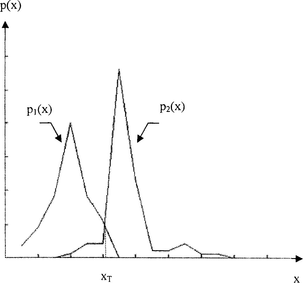
[0028] The following description will assume that the image contains a dark target and a bright background. Segmenting with a threshold T can divide pixels with a gray value less than or equal to the T value and pixels greater than the T value into the target and the background, respectively, and obtain a binary image . Correspondingly, in a binary image, a pixel value of 0 corresponds to the object, and a pixel value of 1 corresponds to the background. Here 1 and 0 are logical values. Of course, the present invention is equally applicable to the case of bright objects and dark backgrounds.

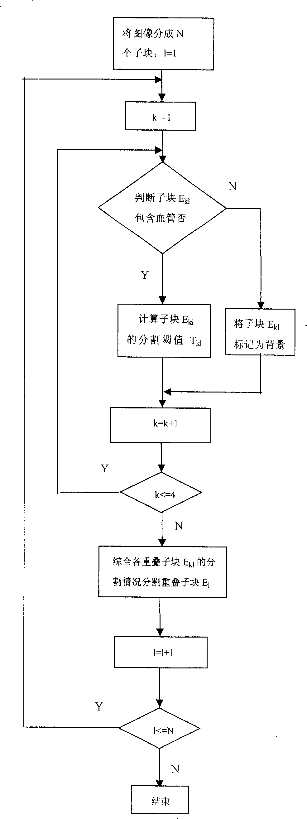
[0029] Combine below figure 1 Describe the steps of the present invention in detail:

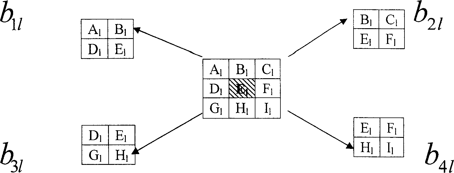
[0030](1) Divide the DSA image into N non-overlapping a1×a2 sub-blocks E l , l=1, 2...N, where, d≤a1≤2d, d≤a2≤2d, where d is assumed to be the maximum value of vessel diameter, the size is b1×b...


PUM


Abstract

A method for dividing blood vessel data in angiogram image of digital imaging includes dividing DSA image to be N numbers of sub block E1 being not superposed; judging whether blood vessel is contained in each of b11, b21, b31 and b41 regions containing sub block E1 or not, dividing superposed sub block in each region, setting one to be equal to one plus one and repeating said judging step and said dividing step on sub block E1 to finalize division on a complete frame of DSA image.

Description

technical field [0001] The invention belongs to the technical field of medical imaging, and in particular relates to a method for segmenting blood vessel data in digital subtraction angiography images. Background technique [0002] Digital Subtraction Angiography (DSA: Digital Subtraction Angiography) technology has been used clinically for more than 20 years, and it is an important basis for non-invasive diagnosis of cardiovascular and cerebrovascular diseases and surgical navigation for interventional therapy. A key task in DSA image processing is to perform image segmentation so that the physiological characteristics of blood vessels can be displayed more clearly, follow-up operations such as structural analysis, motion analysis, and 3D visualization, as well as image-guided surgery, tumor radiation therapy, and treatment evaluation. Applied research is based on image segmentation. Due to the thickness of the tissues that X-rays pass through and the uneven concentration ...

Claims


Application Information

Patent Timeline
Patent Type & Authority Patents(China)
IPC IPC(8): G06T5/00G06K9/34A61B6/00
Inventor 桑农张天序王岳环钟胜李恒彭维雪李艳西
Owner HUAZHONG UNIV OF SCI & TECH
Why Eureka
Try Eureka