
CT image reconstruction method, CT apparatus, and program

a reconstruction method and image technology, applied in the field of ct image reconstruction method and ct apparatus, can solve the problems of data containing inconsistent positional information, affecting the tomographic image, and affecting the accuracy of the interpolation of defective data

Inactive Publication Date: 2009-08-25
GE MEDICAL SYST GLOBAL TECH CO LLC
View PDF10 Cites 2 Cited by
  • Summary
  • Abstract
  • Description
  • Claims
  • Application Information

AI Technical Summary

Benefits of technology

[0011]Therefore, an object of the present invention is to provide a CT image reconstruction method, a CT apparatus, and a program capable of faithfully reflecting the attribute (value and positional information) of substitute data, which is used to compensate defective data, on production of image reconstruction data.
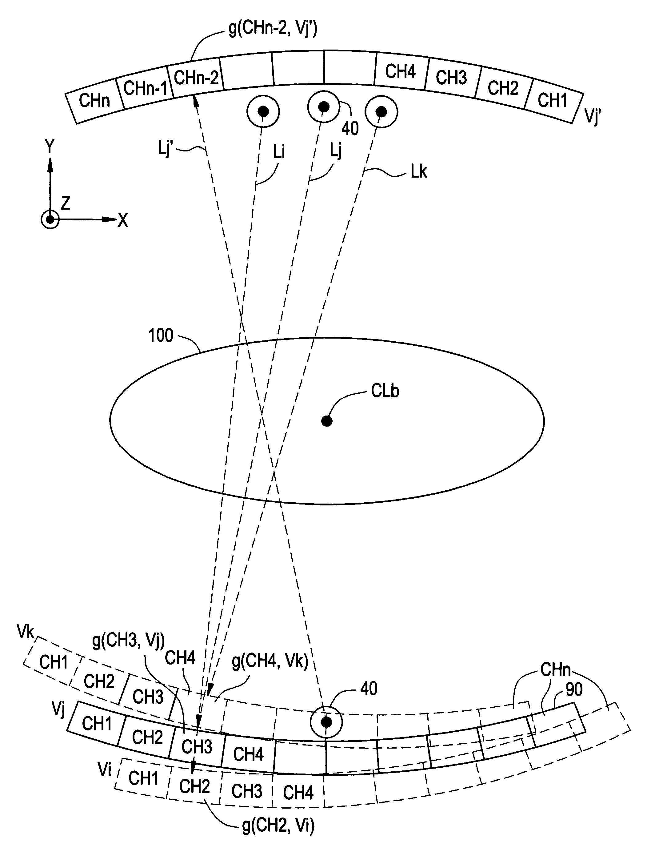
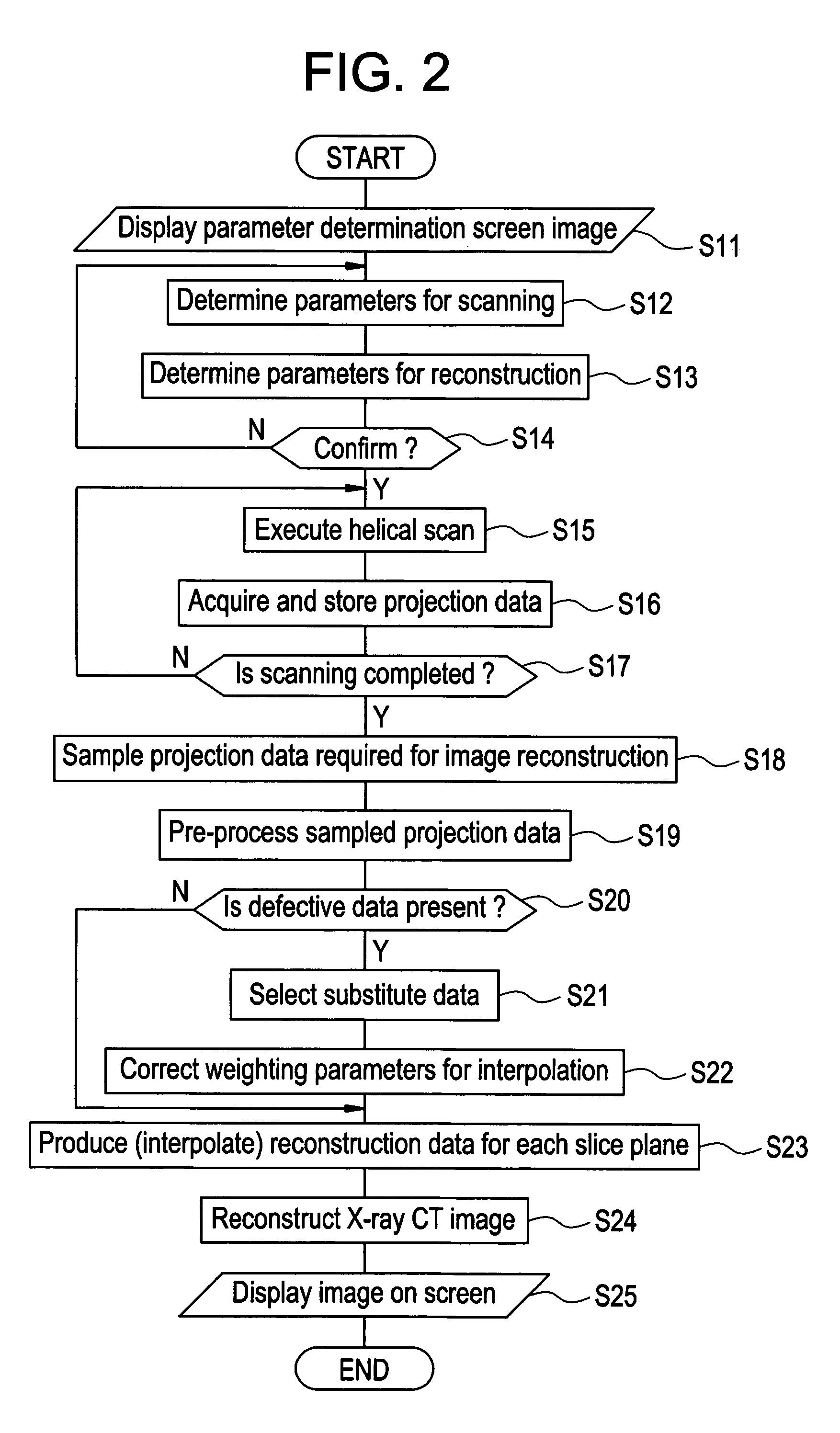
[0012]The above object is accomplished by a technique illustratively shown in FIG. 3. Namely, a CT image reconstruction method according to the first aspect of the present invention comprises the steps of: interpolating back projection data, which is required for reconstruction of a CT image, using projection data items produced by scanning sections of a subject preceding and succeeding a section concerned; and reconstructing a tomographic image of the subject using the interpolated back projection data. Projection data g(CH2,Vi) or g(CH4,Vk) having a predetermined positional relationship to projection data g(CH3,Vj) that is found defective during scanning is substituted for the defective projection data g(CH3,Vj). A value and information on a scanned position (channel and view) contained in the substitute data are used to interpolate back projection data h(CH, Vj). Consequently, the value and scanned position contained in the substitute data are faithfully reflected on interpolation of back projection data. An image is reconstructed more accurately.
[0013]A CT apparatus in accordance with the second aspect of the present invention interpolates back projection data, which is required for reconstruction of a CT image, using projection data items that are produced by scanning sections of a subject preceding and succeeding a section concerned, and reconstructs a tomographic image of the subject according to the interpolated back projection data. The CT apparatus comprises: a data substitution means for substituting projection data, which has a predetermined positional relationship to projection data found defective during scanning, for the defective projection data; and a data interpolation means for interpolating back projection data using a value and information on a scanned position which are contained in the substitute data. Consequently, the value and scanned position contained in the substitute data are faithfully reflected on interpolation of back projection data. Eventually, an image can be reconstructed more accurately.
[0017]According to the sixth aspect of the present invention, the data interpolation means included in the CT apparatus in accordance with the third or fourth aspect thereof interpolates back projection data h(CH3,Vj) by calculating a weighted mean of projection data items g(CH4,Vk) and g(CH3,Vj′), which are produced by scanning sections of a subject, which precede or succeed a section Si in a body-axis direction, at substantially identical view angles Vk and Vj′, as a predetermined function inversely proportional to or based on a distance c or b from a position represented by the interpolated data. Consequently, the value of the substitute data g(CH4,Vk) and the distance c to the position that are contained in the interpolated data are faithfully reflected. Eventually, a tomographic image can be reconstructed more accurately.
[0019]According to the eighth aspect of the present invention, projection data found defective according to the second aspect thereof is projection data whose value is so small as to fall below a predetermined threshold among all projection data items constituting a certain view. Consequently, the poor sensitivity of a detector element can be effectively detected.
[0021]As mentioned above, according to the present invention, a CT image little affected by an artifact and expressing detected data more precisely than a conventional one can be produced.

Problems solved by technology

If defective data is used to reconstruct an image, a tomographic image is affected by a ring artifact or the like.
According to the technique of interpolating defective data itself, when back projection data representing each slice plane whose image is reconstructed is interpolated, pieces of positional information (channel numbers and view angles) on normal projection data items used to interpolate the defective data are lost.
Therefore, the back projection data contains inconsistent positional information.
This deteriorates the quality (faithfulness) of a reconstructed image.

Method used


Image

  • CT image reconstruction method, CT apparatus, and program
  • CT image reconstruction method, CT apparatus, and program
  • CT image reconstruction method, CT apparatus, and program

Examples


Embodiment Construction

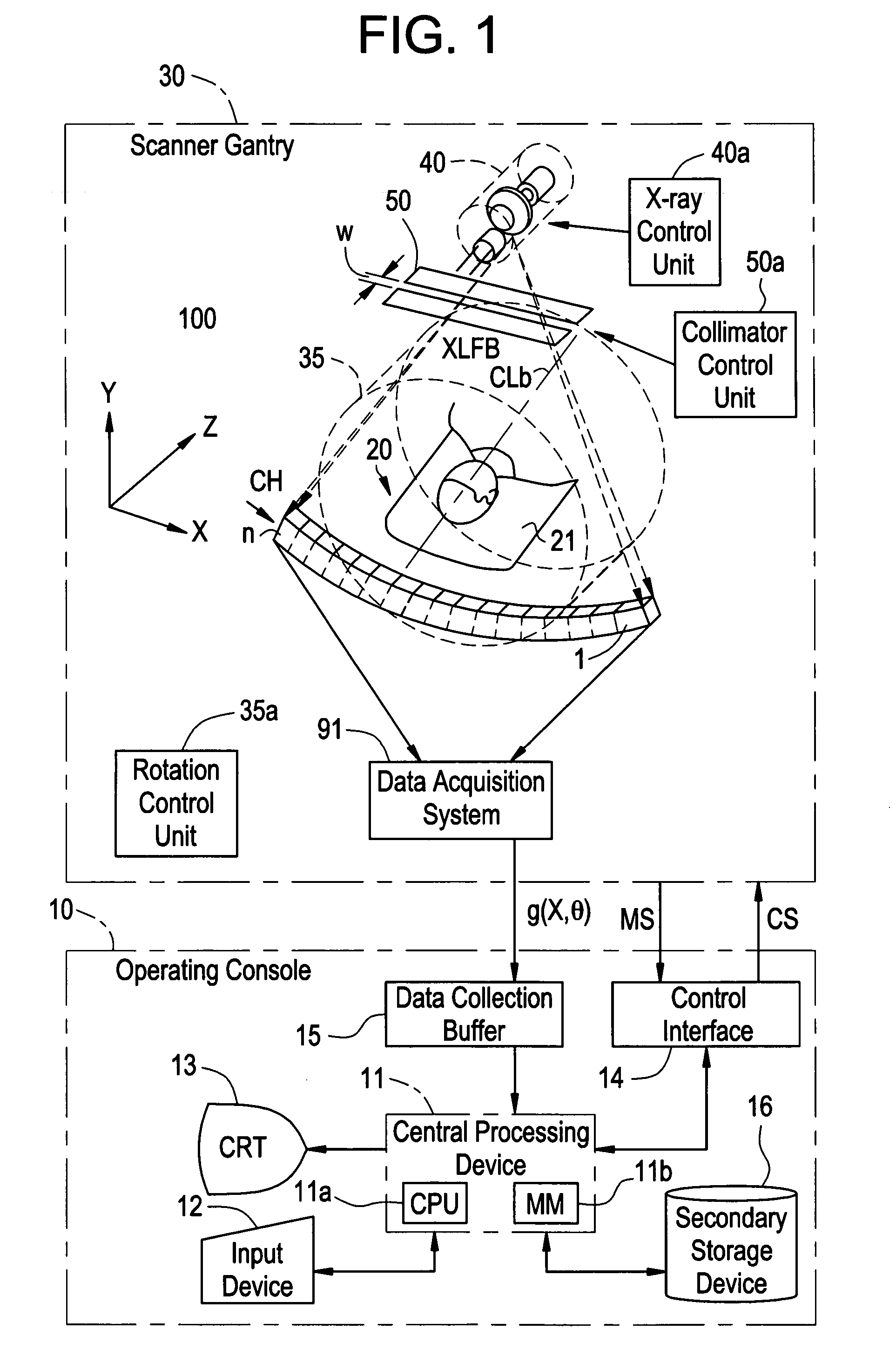
[0027]A preferred embodiment of the present invention will be described below in conjunction with appended drawings. In all the drawings, the same reference numerals denote the same or equivalent components. FIG. 1 shows the configuration of a major portion of an X-ray CT apparatus in accordance with the embodiment. The X-ray CT apparatus comprises a scanner gantry 30 that scans a subject 100 with an X-ray fan-shaped beam XLFB and interprets data, a radiographic table 20 that carries the subject 100 in the directions of a body axis CLb, and an operator console 10 that remotely controls the gantry 30 and table 20 and which an operator or the like manipulates.

[0028]In relation to the scanner gantry 30, reference numeral 40 denotes a rotating anode X-ray tube, and reference numeral 40A denotes an X-ray control unit. Reference numeral 50 denotes a collimator that limits a slice width that is the width of X-rays in a body-axis direction, and reference numeral 50A denotes a collimator con...


PUM


Abstract

A CT image reconstruction method includes substituting other projection data, which has a predetermined positional relationship to projection data that is found defective during scanning, for the defective data, and interpolating back projection data using a value and information on a scanned position that are contained in the substitute data.

Description

CROSS REFERENCE TO RELATED APPLICATIONS[0001]This application claims the benefit of Japanese Application No. 2004-254536 filed Sep. 1, 2004.BACKGROUND OF THE INVENTION[0002]The present invention relates to a CT image reconstruction method, a CT apparatus, and a program. More particularly, the present invention relates to a CT image reconstruction method, a CT apparatus, and a program for interpolating back projection data, which represents each slice plane, using projection data items produced by scanning a subject, and reconstructing a CT image on the basis of the back projection data.[0003]In X-ray CT apparatus, a subject is scanned with an X-ray fan-shaped beam, and an X-ray detector having numerous (for example, 1000) detector elements set in array detects X-rays transmitted by the subject. The subject is intermittently or continuously moved in the directions of the body axis according to an axial or helical scan technique, whereby all projection data items representing a specif...

Claims


Application Information

Patent Timeline
Patent Type & Authority Patents(United States)
IPC IPC(8): A61B6/03
CPCG06T11/005A61B6/027G06T2211/421
Inventor HAGIWARA, AKIRAGOHNO, MAKOTOYAHATA, MITSURU
Owner GE MEDICAL SYST GLOBAL TECH CO LLC
Learn More