
Spatial profiling of proteins using hydrophobic moments

a technology of hydrophobic moments and protein structure, applied in the field of mathematical analysis of proteins, can solve the problems of fewer than these techniques can accurately determine whether a man-made protein structure is or could be a real protein, and thousands of proteins fold, etc., to achieve good mathematical determination, easy to determine, and better quantitative comparison of proteins

Inactive Publication Date: 2008-03-11
IBM CORP
View PDF0 Cites 8 Cited by
  • Summary
  • Abstract
  • Description
  • Claims
  • Application Information

AI Technical Summary

Benefits of technology

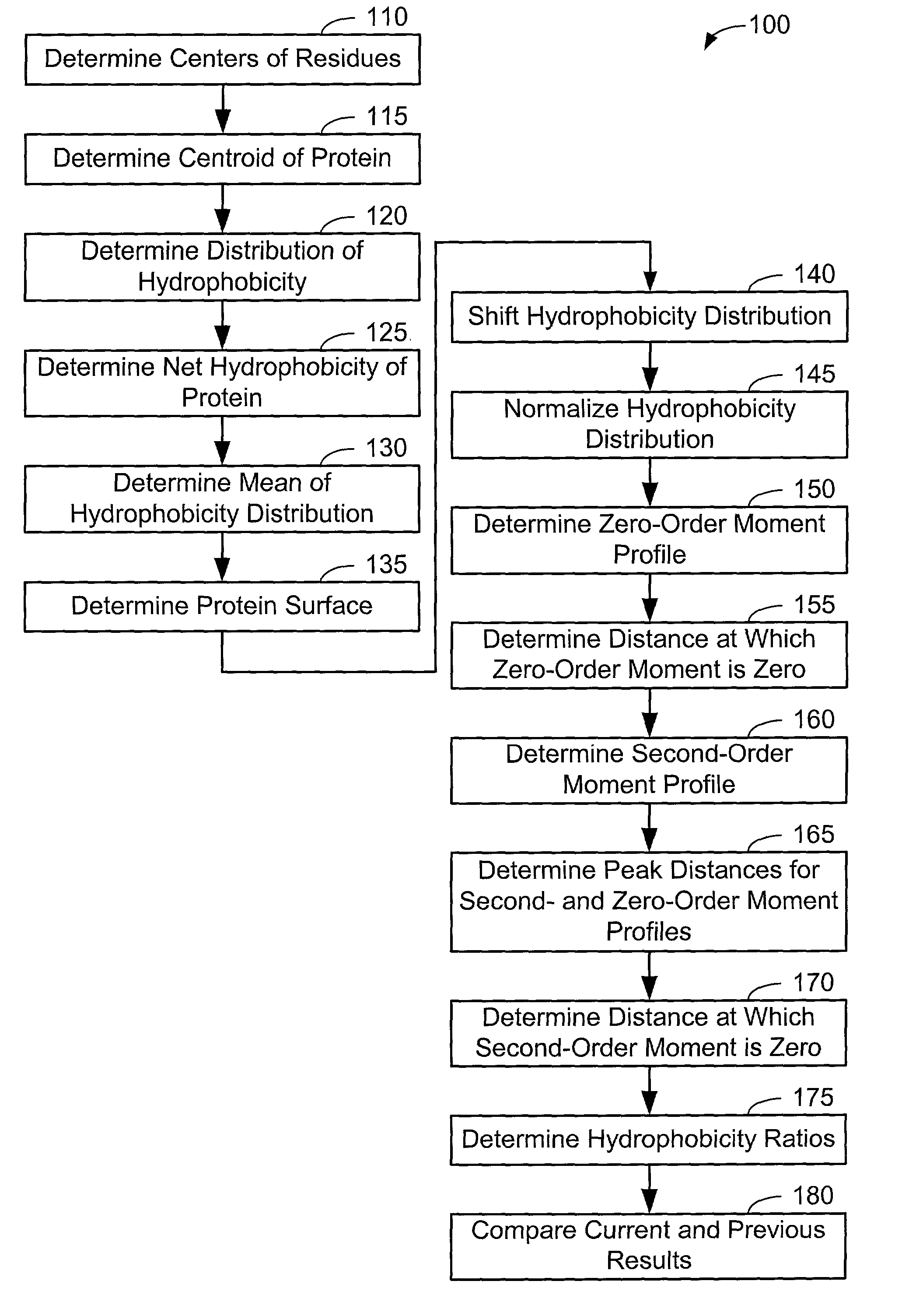
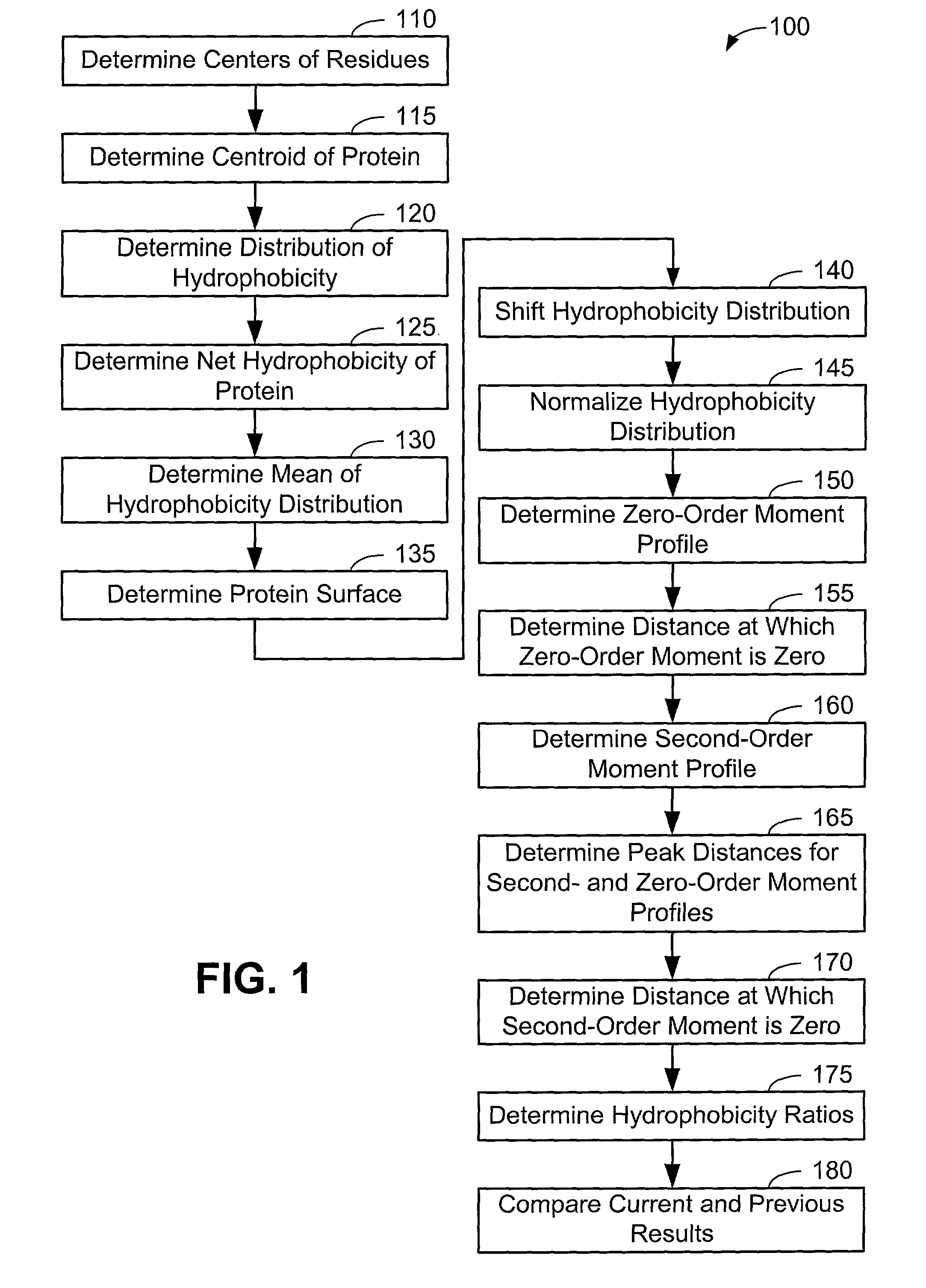
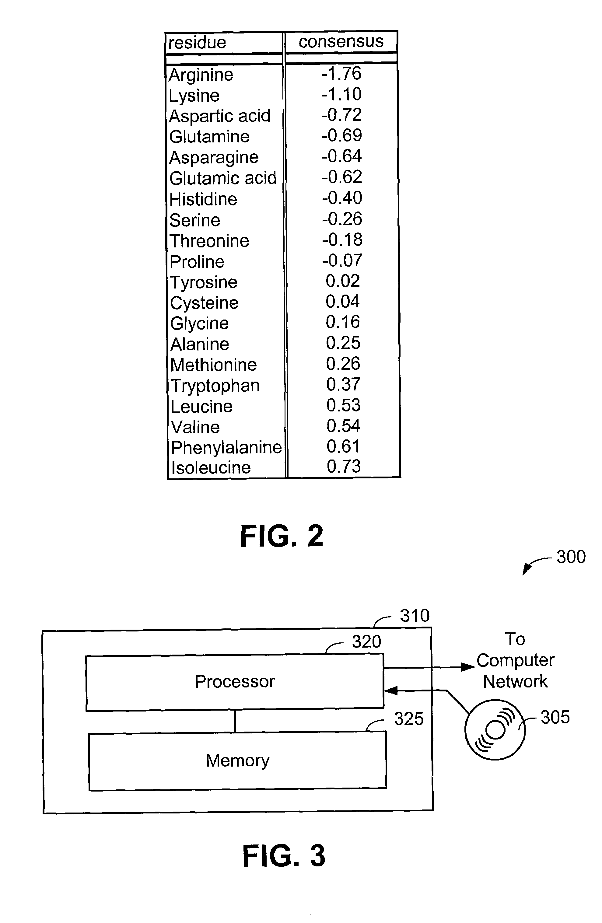
[0008]Generally, the present invention provides a number of procedures to spatially profile proteins by using hydrophobic moments. In all procedures, a hydrophobicity distribution of a protein is shifted and normalized. This allows better quantitative comparisons of proteins. In one procedure, a shape or profile of a curve of a second-order moment of hydrophobicity is determined. This shape can then be used to determine if an example protein belongs to a particular class of proteins, such as globular proteins. A second procedure involves determining one or more ratios, such as the ratio of a distance at which the second order moment of hydrophobicity vanishes to the distance at which a zero-order moment of hydrophobicity vanishes. The distance at which a peak occurs in a profile of the zero- or second-order moment of hydrophobicity can also be used for comparison. These techniques also help to determine if a protein belongs to a globular or other class of proteins. For many of these techniques, a surface or profiling contour can be chosen and used to accumulate hydrophobicities and to determine the moments. These procedures can be combined to provide a good mathematical determination of whether a protein belongs to a particular class of proteins. For globular proteins in particular, the present invention reveals that many globular proteins exhibit similar structural characteristics. This result may be used to easily determine if a decoy protein (a man-made exemplary protein) is a globular protein or a poor structural imitation.

Problems solved by technology

An important problem is determining how hundreds of thousands of proteins fold.
Few of these techniques are suitable for analyzing an entire protein.
Even fewer of the these techniques can accurately determine whether a man-made protein structure is or could be a real protein.

Method used


Image

  • Spatial profiling of proteins using hydrophobic moments
  • Spatial profiling of proteins using hydrophobic moments
  • Spatial profiling of proteins using hydrophobic moments

Examples


examples

[0063]Now that the methods of the present invention have been presented, experimental results will be presented. For the experimental results, protein structures were selected by keyword searches of the Protein Data Bank (PDB) and by examination of entries in different SCOP classes. For more discussion on the latter, see Murzin et al., Journal of Molecular Biology 247, 536-540, 1995, the disclosure of which is incorporated herein by reference. The objective was to choose a selection representative of different sizes and different classes. Thirty protein structures were chosen in this manner. For an internal check, two of the proteins chosen included 1CTQ and 121P, the same protein with independently determined structures. Three additional proteins were also chosen from the recently determined structure of the 30S ribosomal subunit. For more information about the structure of the 30S ribosomal subunit, see Wimberly et al., Nature 407, 327-339, 2000, the disclosure of which is incorpo...


PUM


Abstract

Generally, the present invention provides a number of procedures to spatially profile proteins by using hydrophobic moments. In all procedures, a hydrophobicity distribution of a protein is shifted and normalized. In one procedure, a shape or profile of a curve of a second-order moment of hydrophobicity is determined. A second procedure involves determining one or more ratios, such as the ratio of a distance at which the second order moment of hydrophobicity vanishes to the distance at which a zero-order moment of hydrophobicity vanishes. The distance at which a peak occurs in a profile of the zero- or second-order moment of hydrophobicity can also be used for comparison. For many of these procedures, a surface or profiling contour can be chosen and used to accumulate hydrophobicities and to determine the moments. These procedures can be combined to provide a good mathematical determination of whether a protein belongs to a particular class of proteins.

Description

CROSS REFERENCE TO RELATED APPLICATIONS[0001]This application claims the benefit of U.S. Provisional Application No. 60 / 245,396, filed Nov. 2, 2000.FIELD OF THE INVENTION[0002]The present invention relates to the mathematical analysis of proteins and, more particularly, relates to the spatial profiling of proteins using hydrophobic moments.BACKGROUND OF THE INVENTION[0003]Proteins may be thought of as string with beads on it. Each bead has a particular color. For many proteins, there are 20 colors, or 20 different beads. The string folds up in a certain way, which means that it ends up with a certain series of folds. When profiling a protein, researchers attempt to determine the order of the colors of the beads and where the beads are in three-dimensional space. These locations are important because all of the bodily functions depend on this three-dimensional structure. An important problem is determining how hundreds of thousands of proteins fold.[0004]Many proteins are globular an...

Claims


Application Information

Patent Timeline
IPC IPC(8): G06F17/11G06F17/50G06F19/00G16B15/20G01N33/53G06G7/48G06G7/58
CPCG06F19/16G16B15/00G16B15/20
Inventor SILVERMAN, BENJAMIN D.
Owner IBM CORP
Learn More