
System and method for computer-aided detection and characterization of diffuse lung disease

Inactive Publication Date: 2007-06-26
CHICAGO UNIV OF
View PDF4 Cites 15 Cited by
  • Summary
  • Abstract
  • Description
  • Claims
  • Application Information

AI Technical Summary

Benefits of technology

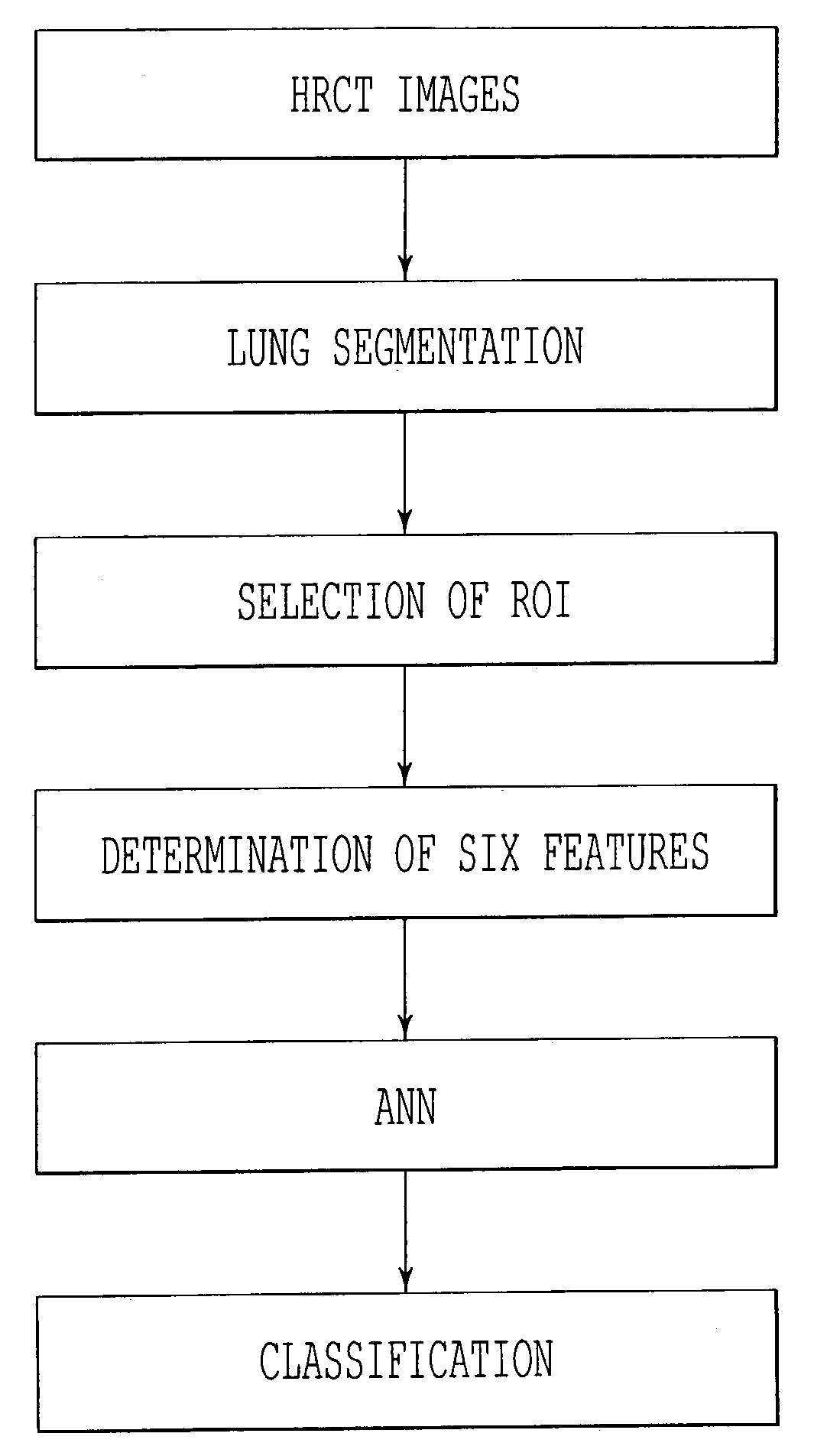
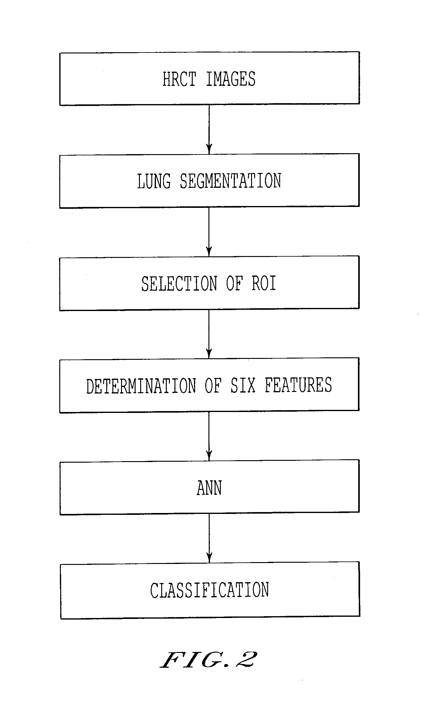
[0027]One object of the present invention is to provide a novel method, system, and computer program product for computer-aided diagnosis of diffuse lung diseases via HRCT in order to assist radiologists' image interpretations. Thus, the present invention is directed to automated computerized detection and characterization of diffuse lung diseases on high-resolution computed tomography (HRCT) images including obtaining image slice data including pixels of an organ; segmenting the image slice data into organ image slice data and non-organ image slice data; extracting predetermined features from the organ image slice data to produce a set of image features; comparing the set of image features against a reference set of organ image features containing image

Problems solved by technology

However, the number of cases used was relatively small, and the level of performance was rather low for clinical applications.
However, it is considered a difficult task for radiologists, partly because of the complexity and variation in diffuse disease patterns on HRCT images, and also because of the subjective terms used for describing diffuse lung diseases.

Method used


Image

  • System and method for computer-aided detection and characterization of diffuse lung disease
  • System and method for computer-aided detection and characterization of diffuse lung disease
  • System and method for computer-aided detection and characterization of diffuse lung disease

Examples


Embodiment Construction

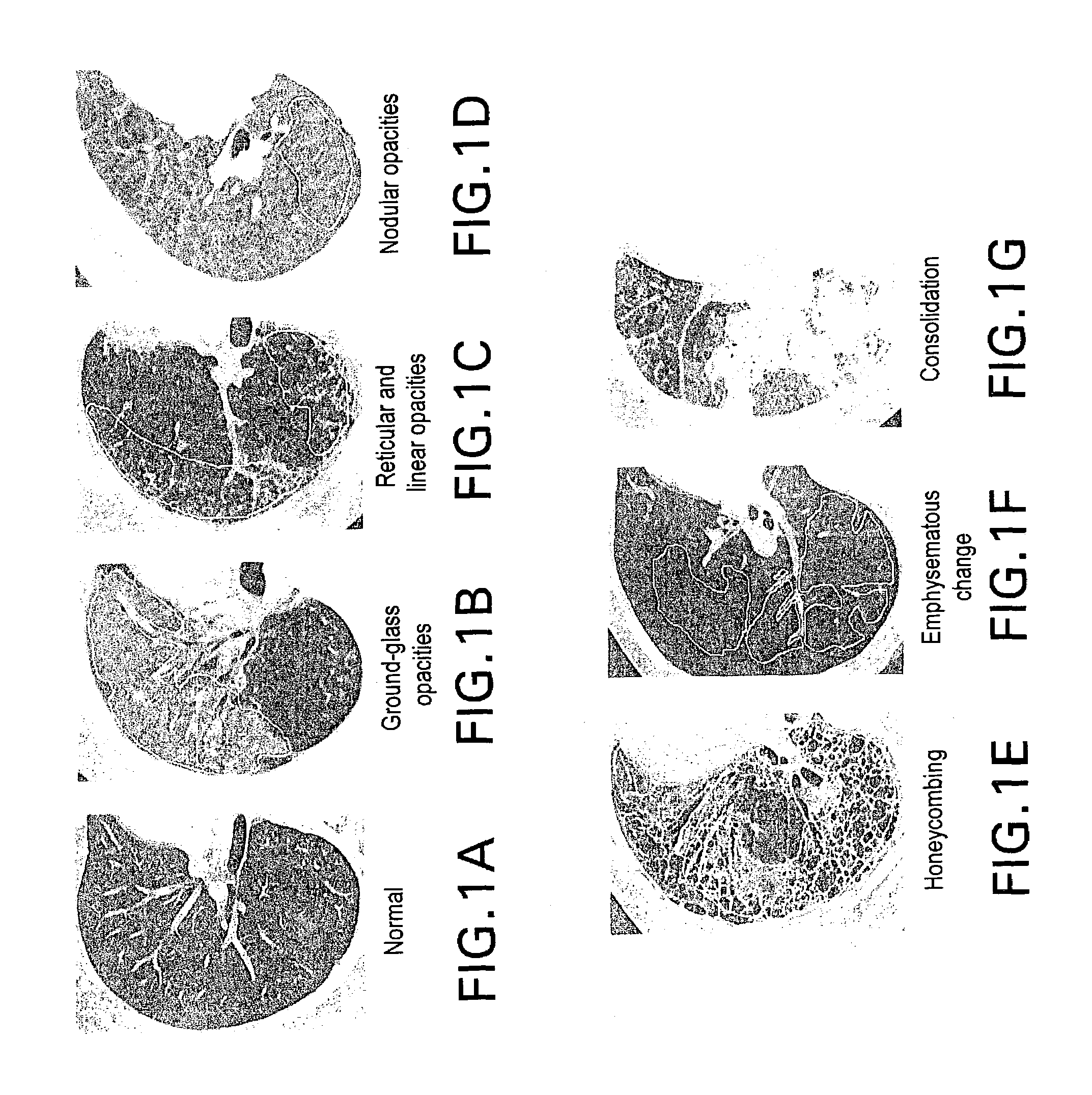
[0041]The present invention uses typical normal and abnormal images as a reference set for diagnostic analysis. The reference set is used because the subjective terms and judgments by radiologists have generally been used to describe diffuse lung diseases. Thus, as prelude to the method of the present invention, an image database of images normal and abnormal cases reported in clinical examinations over a predetermined period of time is examined and representative images are selected as for creation of the reference set. Specific image areas in each image of the selected images are associated with known, specific abnormal patterns and are excerpted and / or otherwise included in the reference set.

[0042]This reference set can be created by chest radiologists independently marking on the reference images areas that are known to include specific diffuse patterns and other abnormalities. This marking may be performed on a CRT monitor and via a mouse. In this way, the chest radiologists ma...


PUM


Abstract

An automated computerized scheme for detection and characterization of diffuse lung diseases on high-resolution computed tomography (HRCT) images including obtaining image data including pixels of an organ; segmenting the image data into organ image data and non-organ image data; extracting predetermined features from the organ image data to produce a set of image features; comparing the set of image features against a reference set of organ image features containing image data known to correspond to normal and abnormal conditions; and producing a comparison result.

Description

[0001]The present invention was made in part with U.S. Government support under USPHS grants CA62625. The U.S. Government may have certain rights to this invention.BACKGROUND OF THE INVENTIONFIELD OF THE INVENTION[0002]This invention relates to an automated computerized scheme for detection and characterization of diffuse lung diseases.[0003]The present invention also generally relates to computerized techniques for automated analysis of digital images, for example, as disclosed in one or more of U.S. Pat. Nos. 4,839,807; 4,841,555; 4,851,984; 4,875,165; 4,907,156; 4,918,534; 5,072,384; 5,133,020; 5,150,292; 5,224,177; 5,289,374; 5,319,549; 5,343,390; 5,359,513; 5,452,367; 5,463,548; 5,491,627; 5,537,485; 5,598,481; 5,622,171; 5,638,458; 5,657,362; 5,666,434; 5,673,332; 5,668,888; 5,732,697; 5,740,268; 5,790,690; 5,832,103; 5,873,824; 5,881,124; 5,931,780; 5,974,165; 5,982,915; 5,984,870; 5,987,345; 6,011,862; 6,058,322; 6,067,373; 6,075,878; 6,078,680; 6,088,473; 6,112,112; 6,138,0...

Claims


Application Information

Patent Timeline
IPC IPC(8): G06K9/00G06T7/00
CPCG06T7/0012G06T2207/30061
Inventor DOI, KUNIOUCHIYAMA, YOSHIKAZUKATSURAGAWA, SHIGEHIKO
Owner CHICAGO UNIV OF
Learn More