
Change detection in medical images

Active Publication Date: 2019-06-20
KONINKLJIJKE PHILIPS NV
View PDF0 Cites 0 Cited by
  • Summary
  • Abstract
  • Description
  • Claims
  • Application Information

AI Technical Summary

Benefits of technology

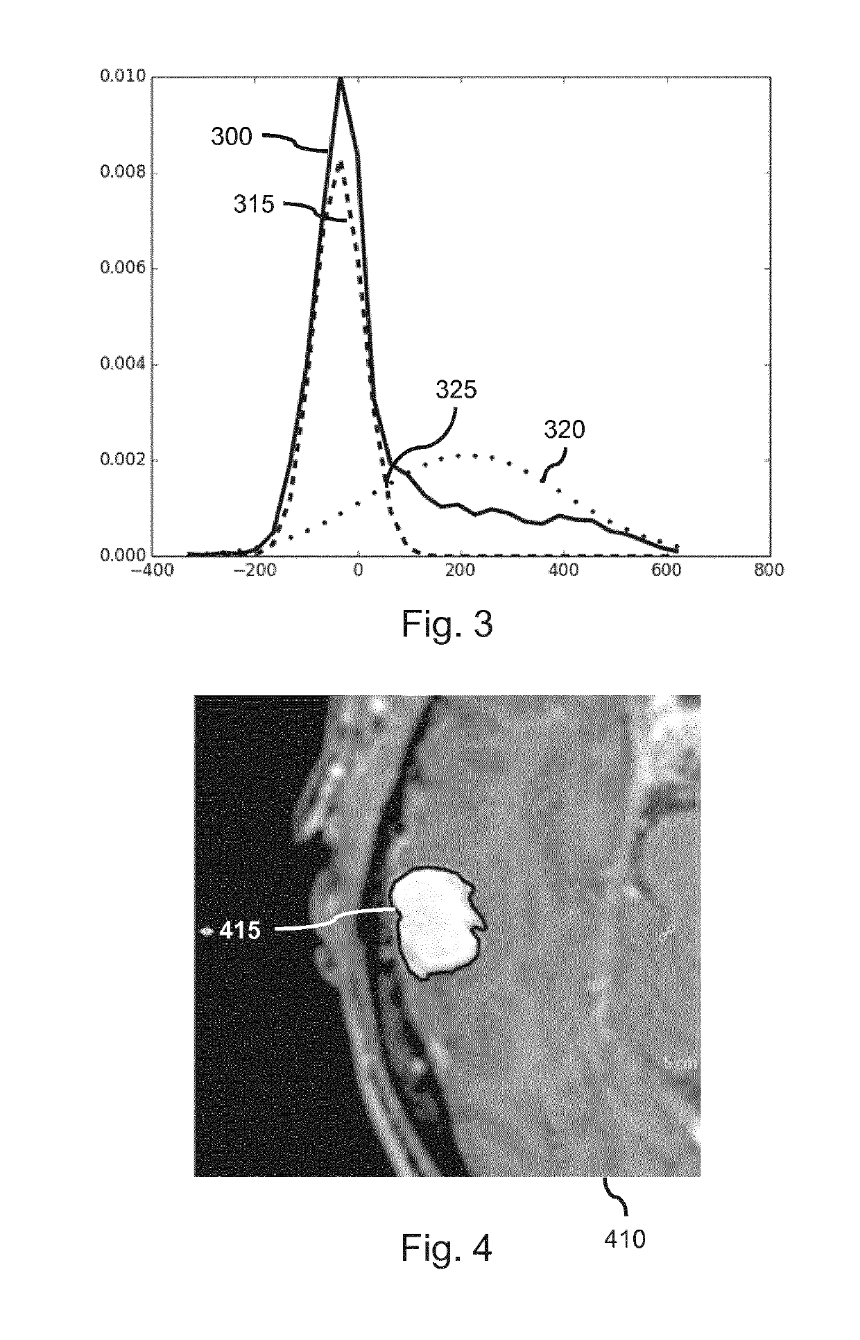
This patent describes a method for detecting changes in medical images by analyzing the distribution of intensities in the images and comparing them to a difference image. The method uses probability distributions and intersection points to accurately detect changes in the images, which can help in the diagnosis and treatment of medical conditions. The method also includes steps to improve the accuracy of the difference image, such as image registration and intensity normalization. Additionally, the method allows for the visualization and evaluation of the detected changes in a user-friendly way, making it easier for medical professionals to analyze and interpret the data.

Problems solved by technology

Disadvantageously, the known approaches to automated change detection are insufficiently accurate.

Method used


Image

  • Change detection in medical images
  • Change detection in medical images
  • Change detection in medical images

Examples


Embodiment Construction

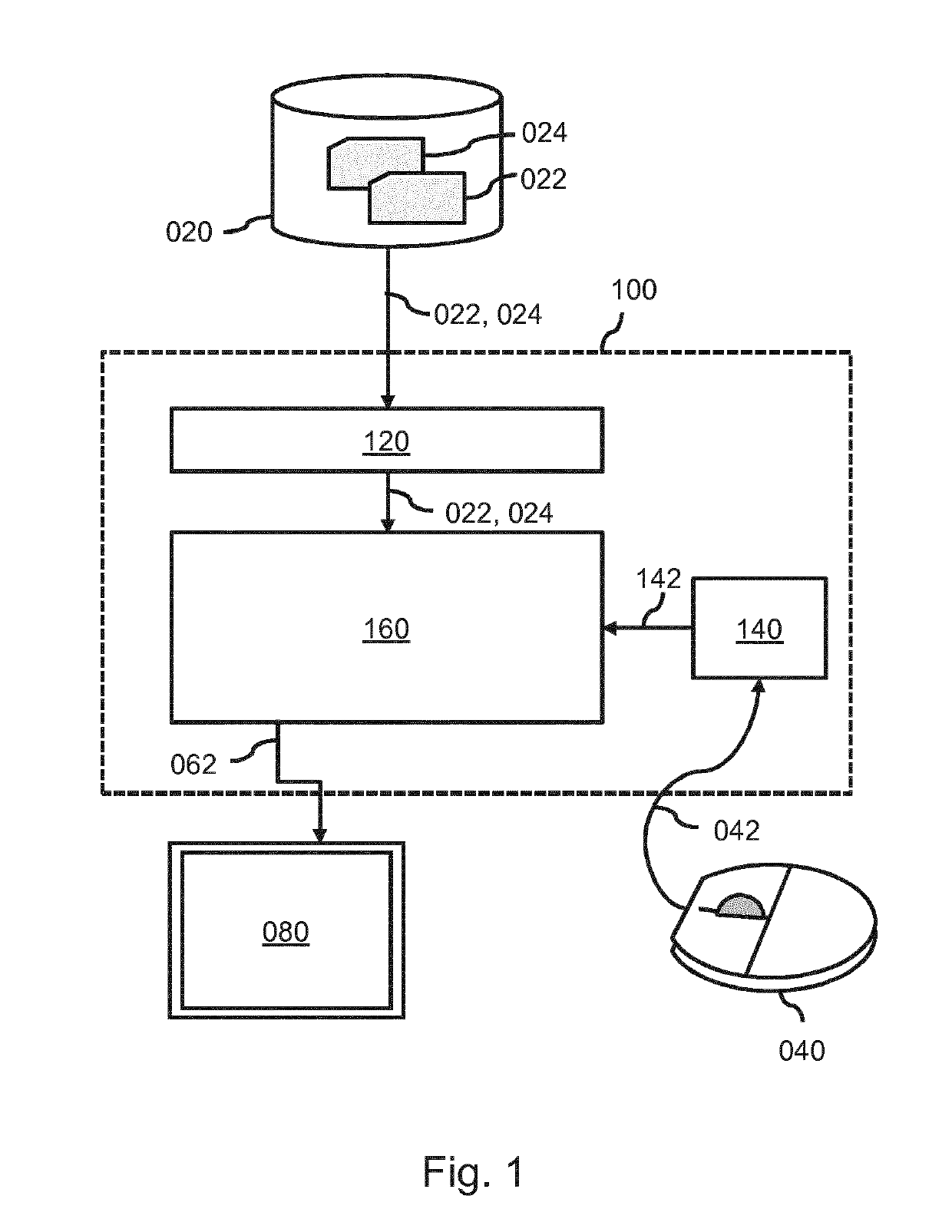
[0087]FIG. 1 shows a system 100 which is configured for change detection in medical images. The system 100 comprises an image data interface 120 configured to access a first medical image and a second medical image. In the example of FIG. 1, the image data interface 120 is shown to be connected to an external image repository 020 which comprises the image data of the first medical image 022 and the second medical image 024. For example, the image repository 020 may be constituted by, or be part of, a Picture Archiving and Communication System (PACS) of a Hospital Information System (HIS) to which the system 100 may be connected or comprised in. Accordingly, the system 100 may obtain access to the image data of the first medical image 022 and the second medical image 024 via the HIS. Alternatively, the image data of the first medical image and the second medical image may be accessed from an internal data storage of the system 100. In general, the image data interface 120 may take va...


PUM


Abstract

A system and method are provided for change detection in medical images. A difference image representing intensity differences between a first medical image and a second medical image is generated. A mixture model is fitted to an intensity distribution of the difference image to identify a plurality of probability distributions which collectively model the intensity distribution. A plurality of intensity ranges is determined as a function of the plurality of probability distributions. Image data of the difference image is labeled by determining into which of the plurality of intensity ranges said labeled image data falls. Accordingly, more accurate change detection is obtained than known systems and method.

Description

FIELD OF THE INVENTION[0001]The invention relates to a system and a method for change detection in medical images. The invention further relates to a server, imaging apparatus and workstation comprising the system. The invention further relates to a computer readable medium comprising instructions to cause a processor system to perform the method.BACKGROUND OF THE INVENTION[0002]Medical images may show an anatomical structure of a patient and / or functional properties of the underlying tissue. It may be desirable to detect changes in (part of) the anatomical structure of the patient, or in the functional properties of the underlying tissue. Such changes may denote a change in disease state or other type of anatomical change. For example, a change may be due to, or associated with, growth of a tumor, progression of Multiple Sclerosis (MS), etc. By detecting the change and the type of change, it may be possible to better treat the disease, e.g., by adjusting a treatment strategy. For t...

Claims


Application Information

Patent Timeline
IPC IPC(8): G06T7/00
CPCG06T7/0016G06T2207/20224G06T2207/20076G06T7/136G16H30/00
Inventor SCHIRMAN, TAMAR DEBORAYEHEZKELY, SHELLY THEODORAKAM, YOSSISHAKIRIN, GEORGYTHIELE, FRANK OLAFKATZ, RUTH
Owner KONINKLJIJKE PHILIPS NV
Learn More