Method and system for spectral characterization in computed tomography x-ray microscopy system

a computed tomography and microscopy technology, applied in the field of spectral characterization in computed tomography x-ray microscopy system, can solve the problems of inconvenient x-ray source spectrum measurement/calibration, time-consuming, and inconvenient x-ray source spectrum calibration, so as to save time, simplify spectrum measurement, and reduce the effect of spectrum measuremen

Active Publication Date: 2017-09-28
CARL ZEISS X RAY MICROSCOPY
View PDF22 Cites 4 Cited by
  • Summary
  • Abstract
  • Description
  • Claims
  • Application Information

AI Technical Summary

Benefits of technology

The present invention proposes a new spectrum measurement and calibration method for x-ray CT systems that has several advantages over current methods. First, it does not require a standard step wedge phantom or cone phantom, which are necessary in current methods. Second, it does not require brute-force measurement of all the attenuation curves for different combinations of x-ray source acceleration voltages, pre-filters, and detectors. Instead, all the attenuation curves can be calculated based on fewer measurements. Third, it reduces the potential for introducing errors due to imperfect alignment of the sample during tomographic reconstructions. Fourth, it simplifies spectrum measurement by eliminating the need for a phantom and reducing the number of spectrum measurements required. Fifth, it saves time by requiring fewer spectrum measurements and eliminating potential calculation errors. Overall, the proposed method simplifies and streamlines the calibration process for x-ray CT systems.

Problems solved by technology

As a result, the x-ray source spectrum measurement and estimation of the various combinations is time consuming.
Further, phantom-based x-ray source spectrum measurement / calibration is not suitable for long-running tomography scans.
This kind of x-ray source spectrum calibration is inconvenient, time-consuming, and can introduce errors in the tomographic reconstructions generated after the sample is replaced and the scan is resumed.
These errors can result if the sample is not positioned precisely into the path of the beam as before the spectral recalibration, in one example.

Method used


Image

  • Method and system for spectral characterization in computed tomography x-ray microscopy system
  • Method and system for spectral characterization in computed tomography x-ray microscopy system
  • Method and system for spectral characterization in computed tomography x-ray microscopy system

Examples


Embodiment Construction

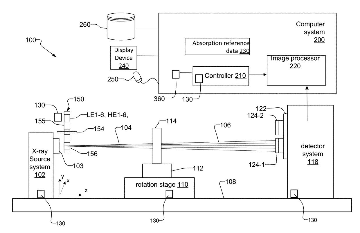
[0053]The invention now will be described more fully hereinafter with reference to the accompanying drawings, in which illustrative embodiments of the invention are shown. This invention may, however, be embodied in many different forms and should not be construed as limited to the embodiments set forth herein; rather, these embodiments are provided so that this disclosure will be thorough and complete, and will fully convey the scope of the invention to those skilled in the art.

[0054]As used herein, the term “and / or” includes any and all combinations of one or more of the associated listed items. Further, the singular forms and the articles “a”, “an” and “the” are intended to include the plural forms as well, unless expressly stated otherwise. It will be further understood that the terms: includes, comprises, including and / or comprising, when used in this specification, specify the presence of stated features, integers, steps, operations, elements, and / or components, but do not pre...


PUM


Abstract

A spectrum measurement and estimation method for tomographic reconstruction, beam hardening correction, dual-energy CT and system diagnosis, etc., comprises determining the spectra for combinations of source acceleration voltage, pre-filters and / or detectors and after measuring the transmission values of several pre-filters, calculating corrected spectra for the combinations of the source acceleration voltage, pre-filters and / or detectors.

Description

RELATED APPLICATIONS[0001]This application claims the benefit under 35 USC 119(e) of U.S. Provisional Application No. 62 / 312,042, filed on Mar. 23, 2016, which is incorporated herein by reference in its entirety.BACKGROUND OF THE INVENTION[0002]X-ray computed tomography (CT) is a non-destructive technique for inspecting and analyzing internal structures of samples. In general, x-rays are absorbed or scattered as the x-rays travel through the sample. The x-rays not absorbed or scattered away are then detected by a detector system. The image formed at the detector system is known as an x-ray projection. Tomographic volume data sets are then reconstructed from a series of these projections at different angles via standard CT reconstruction algorithms such as Filtered Back Projection (FBP).[0003]Some x-ray CT systems utilize polychromatic x-ray sources to generate the x-ray projections. Polychromatic x-ray sources include x-ray tubes (laboratory sources), white synchrotron beams, or acc...

Claims


Application Information

Patent Timeline
IPC IPC(8): G01N23/087G01N23/04
CPCG01N23/087G01N23/046G01N2223/419G01N2223/418G01N2223/309G01N2223/313G01N2223/401G01N2223/3032
Inventor HUANG, ZHIFENGCASE, THOMAS A.STEGER, LOURENS B.
Owner CARL ZEISS X RAY MICROSCOPY
Why Eureka
Try Eureka