Method of Treating Glioma
a glioma and glioma cell technology, applied in the field of glioma treatment, can solve the problems of incomplete understanding of the microenvironmental determinants of glioma cell behavior, high-grade glioma (hggs) is the leading cause of brain tumor death in both children and adults, and achieves the effects of stimulating the proliferation of glial cells, reducing the invasion rate of glioma cells, and reducing the growth rate of hgg
- Summary
- Abstract
- Description
- Claims
- Application Information
AI Technical Summary
Benefits of technology
Problems solved by technology
Method used
Image
Examples
example 1
Optogenetic Control of Cortical Neuronal Activity in a Patient-Derived Pediatric Cortical HGG Orthotopic Xenograft Model
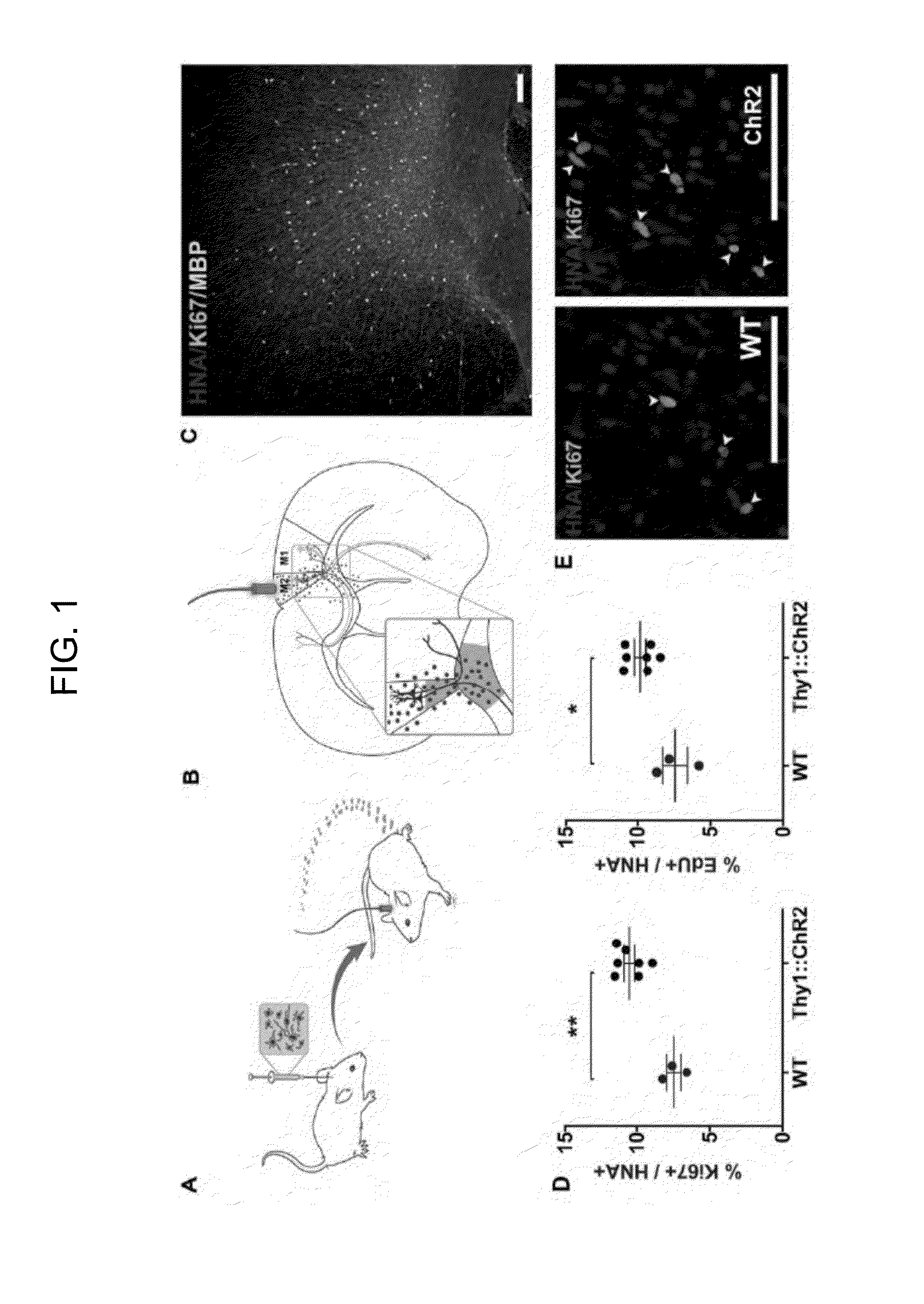
[0220]To test the role of neuronal activity in high-grade glioma (HGG) growth, in vivo optogenetic stimulation of the premotor cortex in awake, freely-behaving mice bearing patient-derived orthotopic xenografts of pediatric cortical glioblastoma was employed (FIG. 1, panels A-C). The well-characterized Thy1::ChR2 mouse model expressing the excitatory opsin channelrhodopsin-2 (ChR2) in deep layer cortical projection neurons was crossed onto an immunodeficient background (NOD-SCIDIL2R γ-chain-deficient, NSG), resulting in a mouse model (Thy1::ChR2;NSG) amenable to both in vivo optogenetic stimulation and orthotopic xenografting. ChR2-expressing neurons respond to pulses of blue (473-nm) light with millisecond precision. This allows for neuronal spiking frequency to be determined by the frequency of light pulses delivered. Expression of ChR2 does not alter the electri...
example 2
Neuronal Activity Promotes Glioma Proliferation Through Secreted Factors
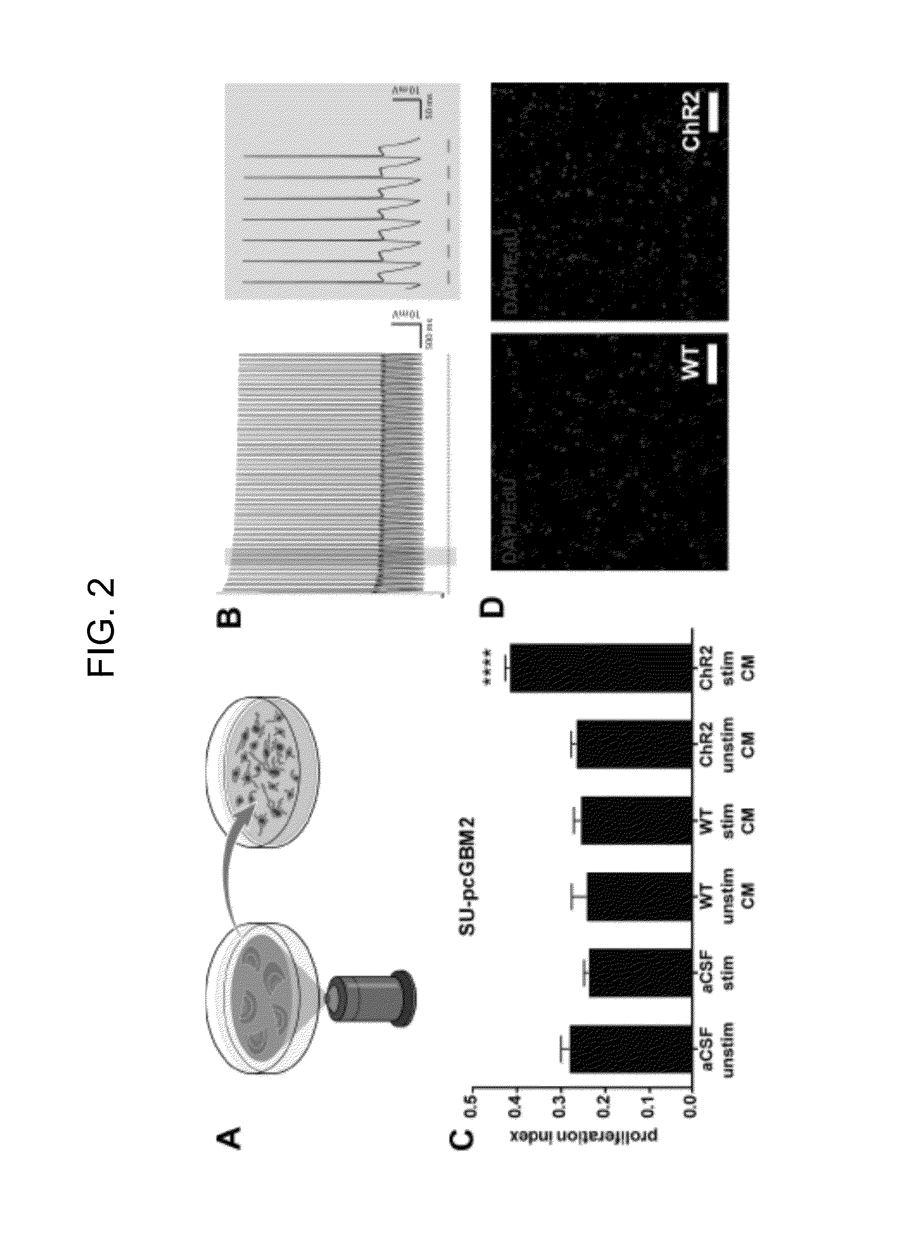
[0228]To determine if neurons stimulate glioma proliferation through secretion of an activity-regulated mitogen or mitogens, acute cortical slices from Thy1::ChR2 or identically-manipulated WT mice were optogenetically stimulated in situ and the conditioned medium (CM) was collected. Patient-derived HGG cultures were then exposed to the collected CM (FIG. 2, panel A). The slice stimulation paradigm mirrored the in vivo paradigm, using 473-nm light at 20 Hz for cycles of 30 seconds on, 90 seconds off over a 30-minute period. For this in situ optogenetic model, the expected neuronal firing response to blue light was validated electrophysiologically, confirming 20-Hz spike trains for 30-second periods throughout the 30-minute stimulation paradigm (FIG. 2, panel B). Maintenance of slice health throughout the stimulation paradigm was confirmed histologically and electrophysiologically (FIG. 9). Cortical slices from W...
example 3
Activity-Regulated Glioma Mitogen(s) are Secreted Proteins
[0249]A series of biochemical analyses were employed to ascertain the nature of the activity-regulated mitogen(s). To determine if the mitogen(s) are small molecules or macromolecules, the CM from optogenetically stimulated Thy1::ChR2 or identically manipulated cortical slices was collected as above and fractionated by molecular size into 10 kDa fractions. The in vitro proliferation assay was repeated and it was found that the >10 kDa macromolecular fraction, but not the 100 degrees Celsius to denature proteins, resulting in loss of the proliferation-inducing capacity of the CM (FIG. 3, panel C). In contrast, treatment of the CM with RNase and DNase had no effect on the mitogenic effect of the CM on glioma cells (FIG. 3, panel D). Taken together, these data indicate that the neuronal activity-regulated secreted mitogen(s) is a protein between 10 and 100 kDa.
[0250]FIG. 3: Cortical Neuronal Activity-Regulated Glioma Mitogen(s) ...
PUM
| Property | Measurement | Unit |
|---|---|---|
| Fraction | aaaaa | aaaaa |
Abstract
Description
Claims
Application Information
 - Generate Ideas
- Intellectual Property
- Life Sciences
- Materials
- Tech Scout
- Unparalleled Data Quality
- Higher Quality Content
- 60% Fewer Hallucinations
Browse by: Latest US Patents, China's latest patents, Technical Efficacy Thesaurus, Application Domain, Technology Topic, Popular Technical Reports.
© 2025 PatSnap. All rights reserved.Legal|Privacy policy|Modern Slavery Act Transparency Statement|Sitemap|About US| Contact US: help@patsnap.com


