
Endoscopic apparatus with integrated hemostasis device

a technology of hemostasis device and endoscope, which is applied in the field of endoscope, can solve the problems of tedious and time-consuming aforementioned procedures, not without its deficiencies,

Inactive Publication Date: 2013-01-03
BOSTON SCI SCIMED INC
View PDF6 Cites 2 Cited by
  • Summary
  • Abstract
  • Description
  • Claims
  • Application Information

AI Technical Summary

Problems solved by technology

While this method may be effective in treating internal bleeding, it is not without its deficiencies.
For example, the aforementioned procedure is tedious and time consuming because of the need to introduce, position, energize, and withdraw the RF probe if other implements, e.g. biopsy forceps, are needed between the treatment of two bleeders.

Method used


Image

  • Endoscopic apparatus with integrated hemostasis device
  • Endoscopic apparatus with integrated hemostasis device
  • Endoscopic apparatus with integrated hemostasis device

Examples


Embodiment Construction

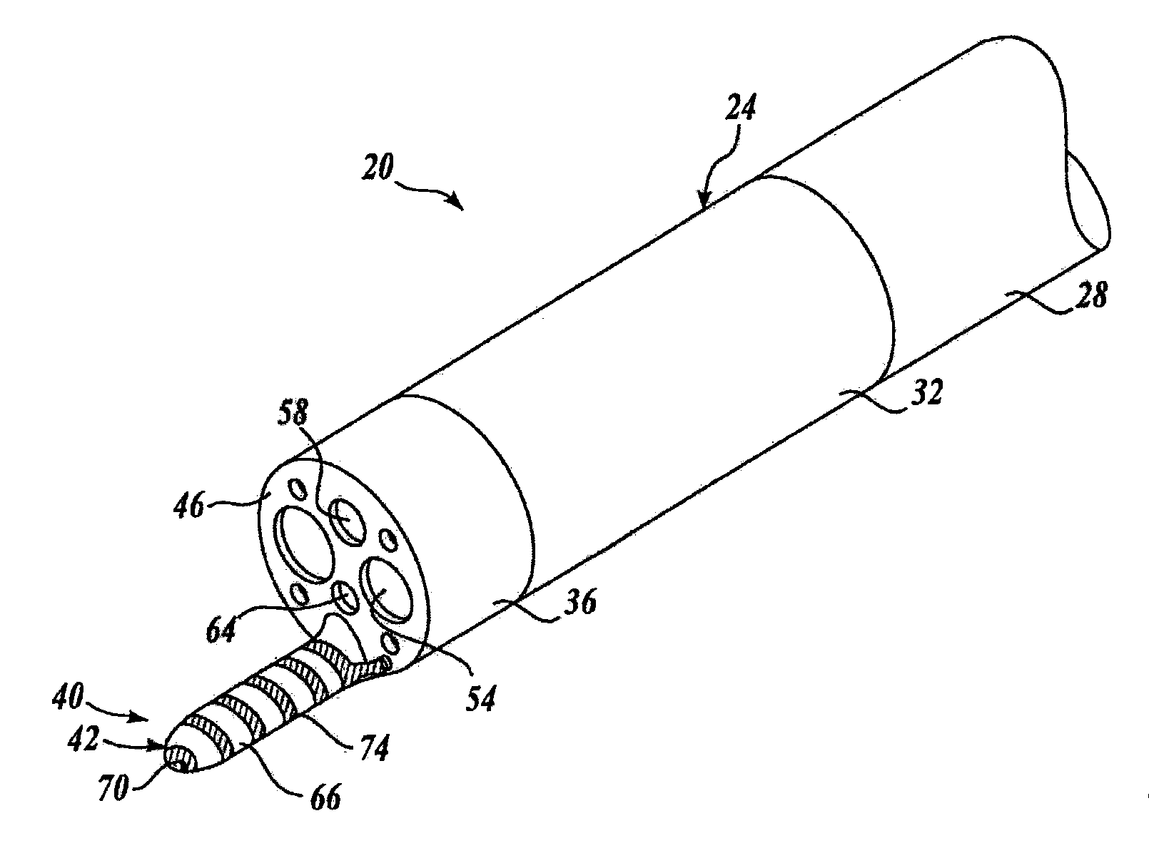
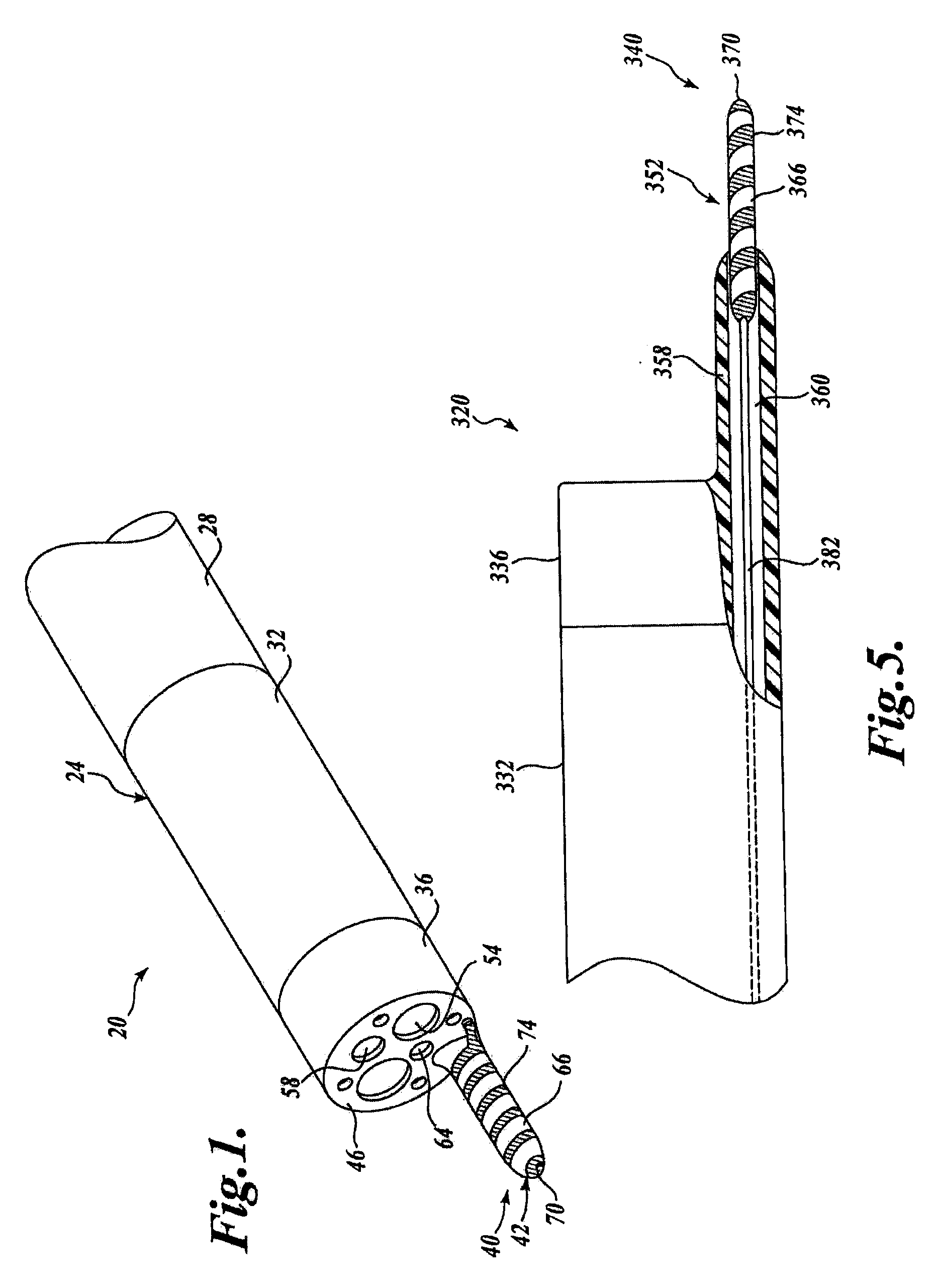
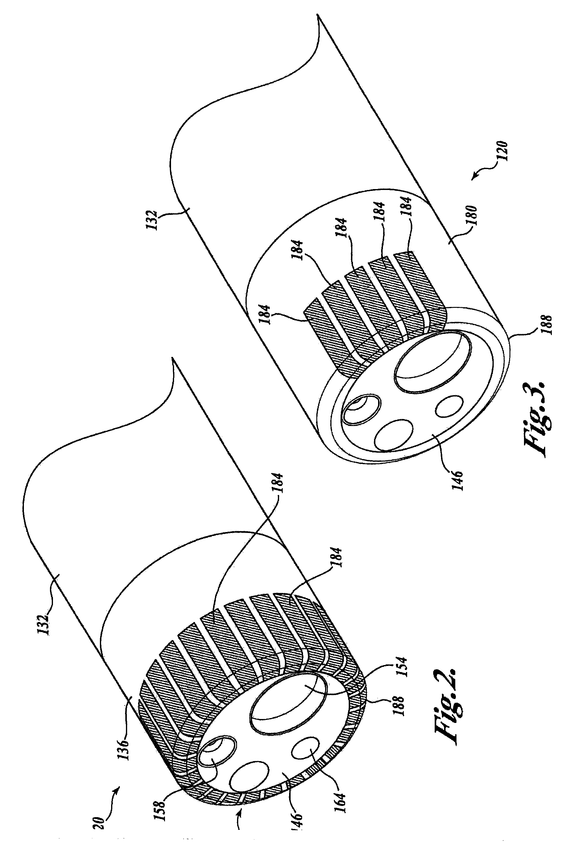
[0016]The present invention will now be described with reference to the drawings where like numerals correspond to like elements. Embodiments of the present invention are directed to devices of the type broadly applicable to numerous medical applications in which it is desirable to insert an imaging device, catheter or similar device into a body lumen or passageway. Specifically, embodiments of the present invention are directed to medical devices having hemostasis capabilities. Several embodiments of the present invention are directed to medical devices having hemostasis capabilities that incorporate endoscopic features, such as illumination and visualization capabilities, for endoscopically viewing anatomical structures within the body. As such, embodiments of the present invention can be used for a variety of different diagnostic and interventional procedures, including colonoscopy, upper endoscopy, bronchoscopy, thoracoscopy, laparoscopy and video endoscopy, etc., and are partic...


PUM


Abstract

An endoscope or other medical device includes an elongated shaft having a flexible proximal section, an articulatable distal region, and distal tip. The endoscope or other device includes an associated hemostasis device for treating internal bleeding during a contemporaneous medial procedure. Embodiments of the present invention may incorporate mechanical, chemical, and / or electrical techniques for performing hemostasis.

Description

FIELD OF THE INVENTION[0001]The present invention is generally directed to endoscopic apparatuses for use in medical procedures, and in particular, to endoscopic apparatuses having associated hemostasis devices.BACKGROUND OF THE INVENTION[0002]It has become well established that there are major public health benefits from regular endoscopic examinations as an aid to the early detection of disease of internal structures such as the alimentary and excretory canals and airways, e.g., the colon, esophagus, lungs, uterus, bladder, bronchi, and other organ systems. A conventional imaging endoscope used for such procedures comprises a flexible tube with a fiber optic light guide that directs illuminating light from an external light source to the distal tip where it illuminates the region (i.e. tissue, occlusion object) to be examined. Frequently, additional optical components are incorporated to adjust the spread of the light exiting the fiber bundle and the distal tip. An objective lens ...

Claims


Application Information

Patent Timeline
Patent Type & Authority Applications(United States)
IPC IPC(8): A61B18/12A61B17/32A61B18/14A61B18/18
CPCA61B1/00087A61B1/018A61B18/1492A61B2018/1495A61B2018/00494A61B2018/00982A61B2018/00482
Inventor COUVILLON, JR., LUCIEN ALFRED
Owner BOSTON SCI SCIMED INC
Learn More