Encapsulated cells and composites thereof
a technology of encapsulated cells and composites, which is applied in the direction of microcapsules, capsule delivery, biocide, etc., can solve the problems of not being able to encapsulate cells directly in it, and not being able to provide a friendly environment for cell survivability, so as to enhance the availability of nutrients for encapsulated cells, easy gelling properties, and increase cell viability
- Summary
- Abstract
- Description
- Claims
- Application Information
AI Technical Summary
Benefits of technology
Problems solved by technology
Method used
Image
Examples
example 1
[0176]This example relates to gel microstructure regulation of proliferation and differentiation of MC3T3-E1 cells encapsulated in alginate beads.
[0177]Materials and Methods: An O / W emulsion technique was used for encapsulation of cells in alginate beads. Alginic acid (viscosity 20,000˜40,000 cps, Aldrich) was dissolved in Di-water and in α-MEM at the concentrations with 1 and 2% (w / v). MC3T3-E1 embryonic mouse osteoblast precursor cells were suspended in the alginate solutions. Cell alginate suspensions were then added dropwise into two kinds of Ca-catalysts: (a) CaCl2 in Di-water and (b) CaCl2 in α-MEM. The concentrations of CaCl2 solutions were 100 and 200 mM. The beads, thus formed, were cured in the Ca-catalyst medium for 2 hr. MC3T3-E1 encapsulated with 1×10⁶ cells / ml in alginate beads. The viability of encapsulated MC3T3-E1 cells was quantified using the live / dead viability assay. Cell differentiation in the alginate beads was characterized using alkaline phosphatase (ALP) an...
example 2
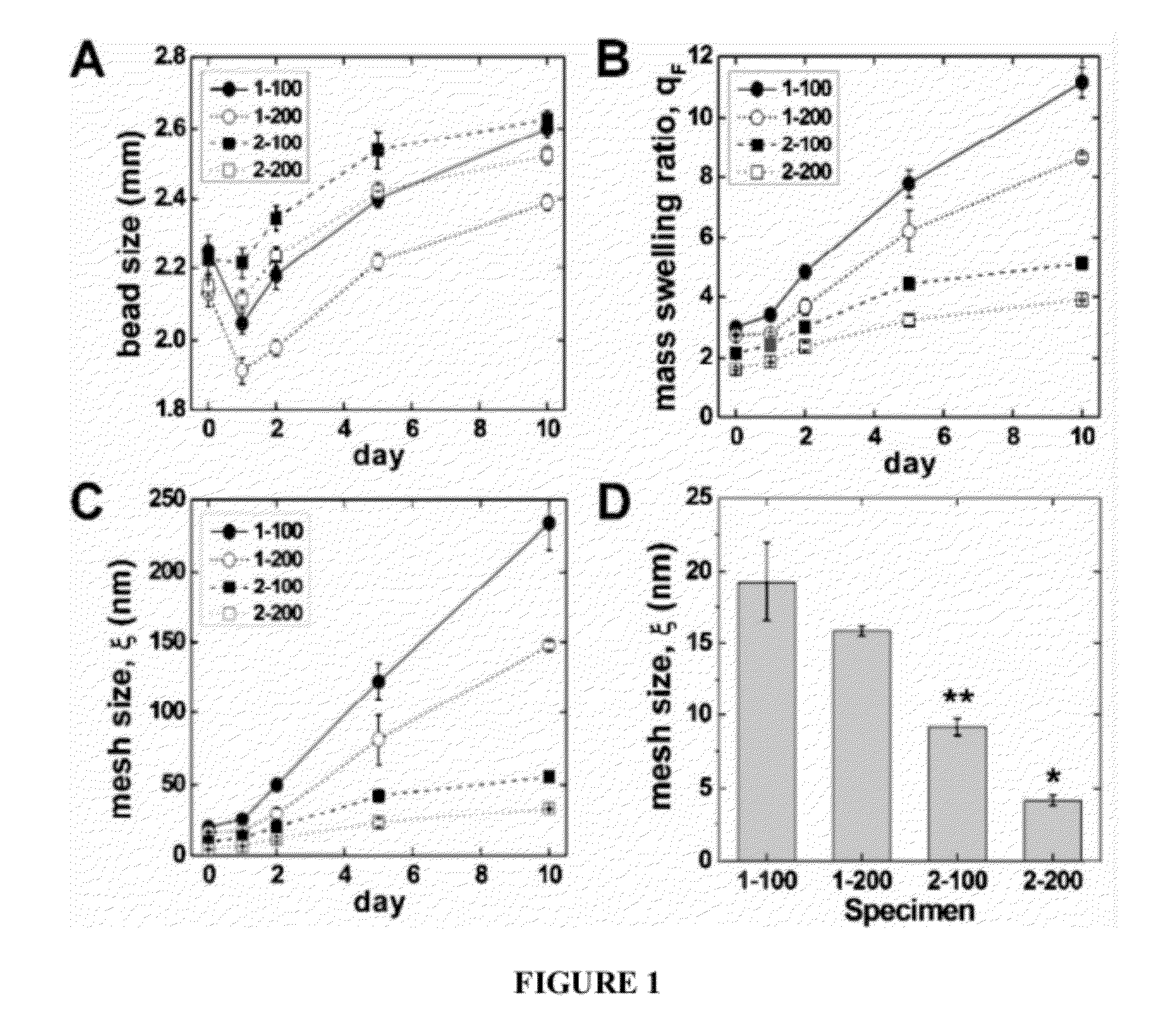
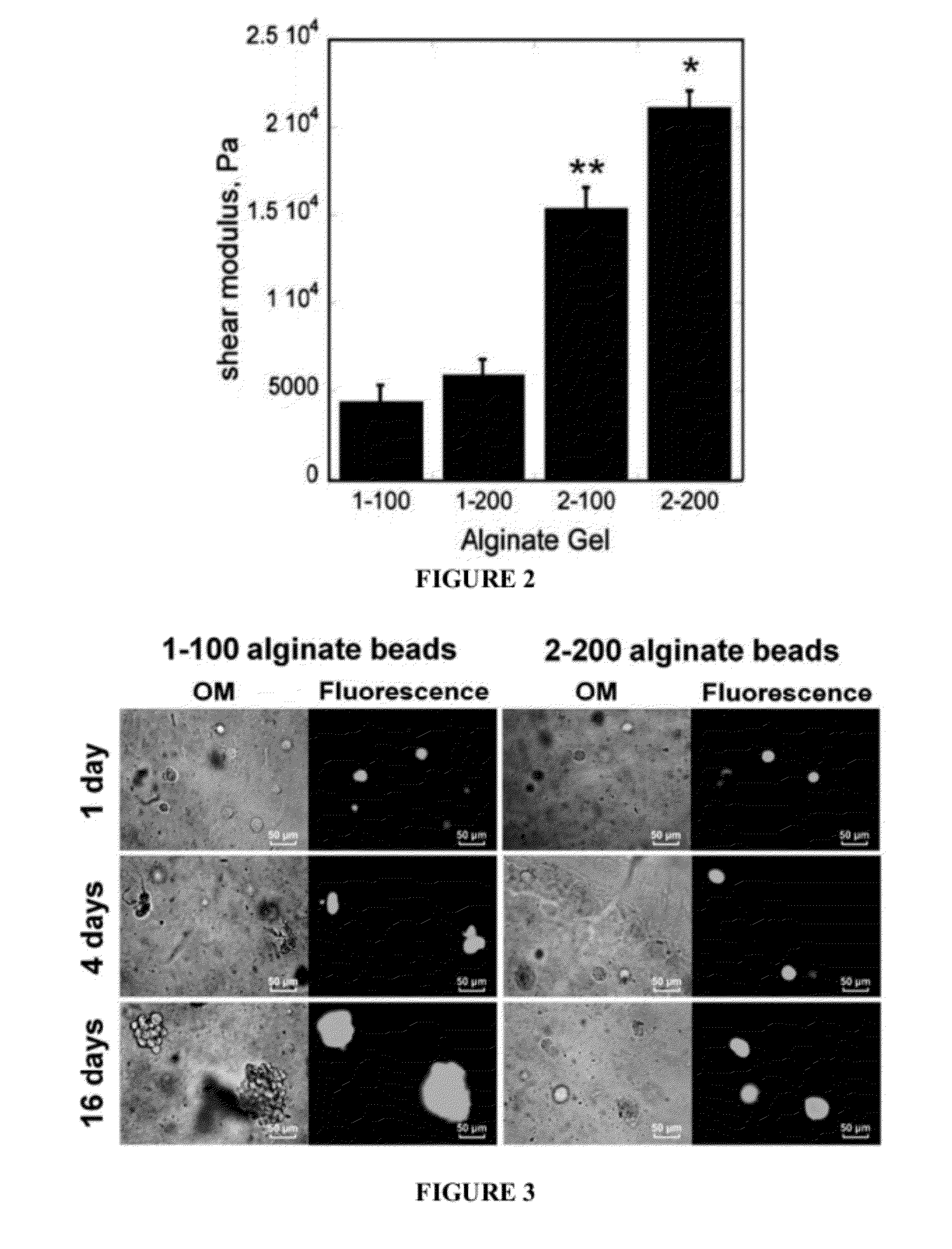
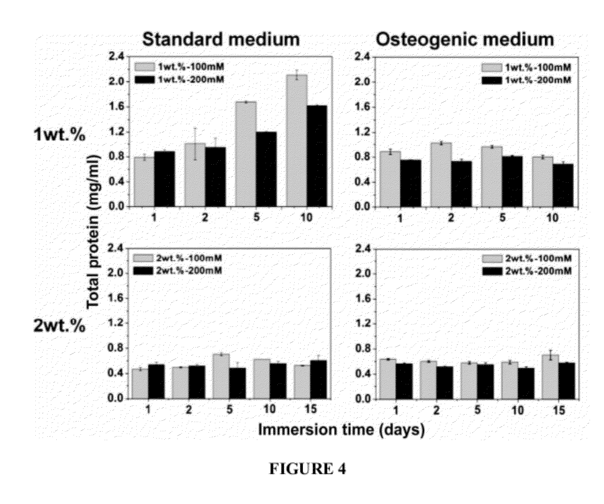
[0180]This Example also relates to the changes in proliferation and differentiation of MC3T3-E1 cells in alginate beads with differing mesh microstructures.
[0181]Materials and Methods
[0182]Alginic acid sodium salt (viscosity 20,000˜40,000 cP, molecular weight 120-190 kDa, Aldrich, St. Louis, Mo.) was dissolved in DI-water and in α-MEM at concentrations of 1 and 2% (w / v). Briefly, MC3T3-E1 cells (1×10⁶ cells / ml) were suspended in the alginic acid sodium salt solutions (1 and 2% (w / v)) and mixed for 2 h. Cell viability was measured by live / dead staining before and after incubation in alginic acid solution and found to be unchanged. Suspensions of cells in alginate were then added drop-wise into two different crosslinker solutions: (a) CaCl2 in DI-water or (b) CaCl2 in α-minimum essential medium (MEM) at room temperature. The concentration of CaCl2 solutions was either 100 or 200 mM. The beads formed in the microencapsulation device were subsequently cured in the CaCl2 solution for 1 h...
example 3
[0202]This example relates to local cell delivery from injectable biodegradable polymeric scaffolds.
[0203]Materials and Methods: Similarly to Examples 1 and 2, an O / W emulsion technique was used for encapsulation of cells in alginate beads. MC3T3-E1 embryonic mouse osteoblast precursor cells were encapsulated with 1×10⁶ cells / ml in alginate beads. Alginate beads were prepared using Ca-catalysts as CaCl2 in α-MEM. The loading of cell-encapsulated beads in the reactive PUR scaffold was 50 wt %. Cell survivability in alginate beads was determined using live / dead staining Cell differentiation in the alginate beads, beads alone, and beads incorporated in PUR, was characterized using alkaline phosphatase (ALP) and osteocalcin.
[0204]Results: Biomimetic cell-alginate capsules are successfully synthesized by O / W emulsion technique. The synthesized Ca-alginate / PUR composite exhibited three different pore structures: macropores (0.5˜2 mm) from degradation of alginate beads, intermediate pores ...
PUM
| Property | Measurement | Unit |
|---|---|---|
| Fraction | aaaaa | aaaaa |
| Fraction | aaaaa | aaaaa |
| Fraction | aaaaa | aaaaa |
Abstract
Description
Claims
Application Information
 - R&D
- Intellectual Property
- Life Sciences
- Materials
- Tech Scout
- Unparalleled Data Quality
- Higher Quality Content
- 60% Fewer Hallucinations
Browse by: Latest US Patents, China's latest patents, Technical Efficacy Thesaurus, Application Domain, Technology Topic, Popular Technical Reports.
© 2025 PatSnap. All rights reserved.Legal|Privacy policy|Modern Slavery Act Transparency Statement|Sitemap|About US| Contact US: help@patsnap.com


