Eureka AIR delivers breakthrough ideas for toughest innovation challenges, trusted by R&D personnel around the world.

Method and System for Virtual Percutaneous Valve Implantation

a technology implantation method, which is applied in the field of virtual percutaneous valve implantation, can solve the problems of repeated valve replacement, high mortality, stroke, and high mortality, and achieve the effects of reducing the risk of strok

Active Publication Date: 2011-06-23
SIEMENS HEALTHCARE GMBH
View PDF9 Cites 66 Cited by
  • Summary
  • Abstract
  • Description
  • Claims
  • Application Information

AI Technical Summary

Benefits of technology

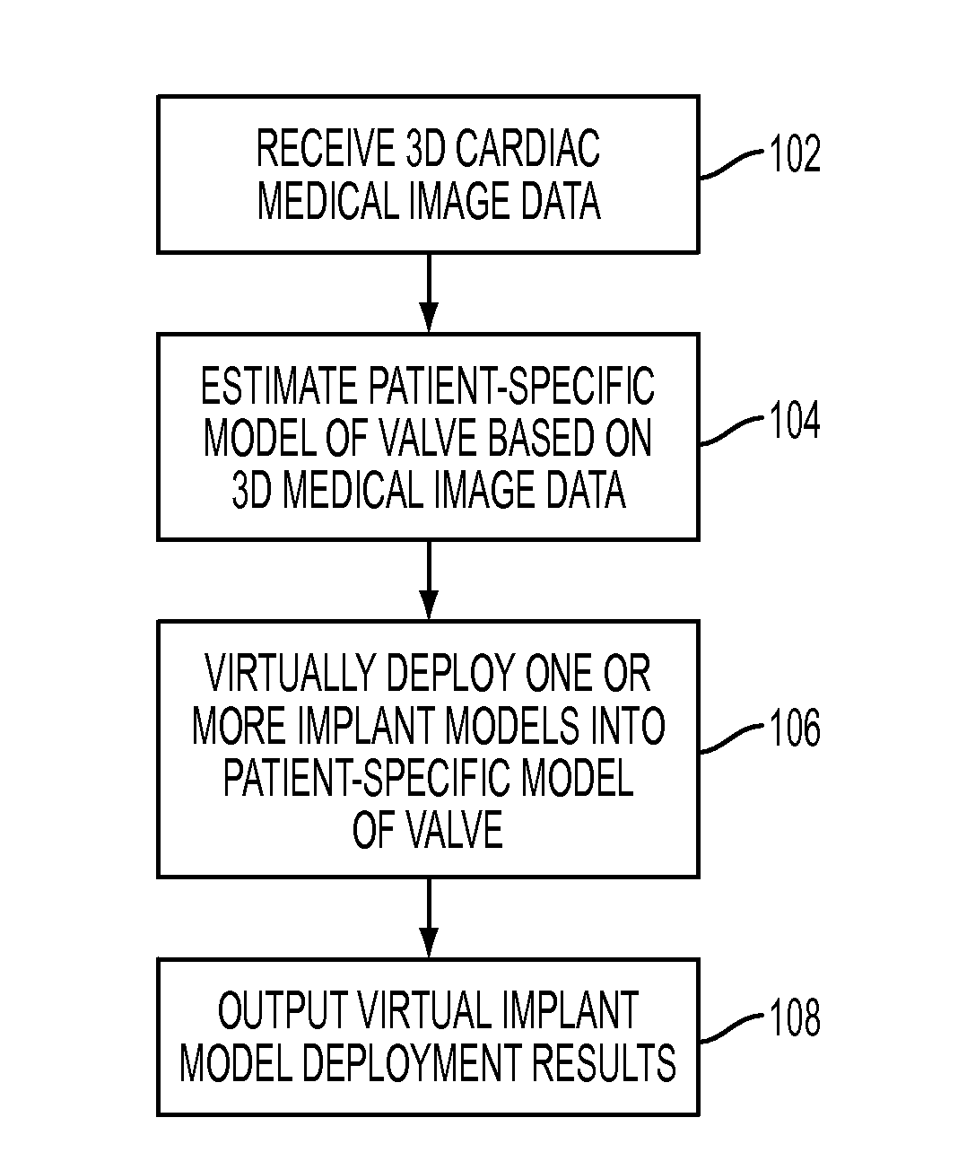
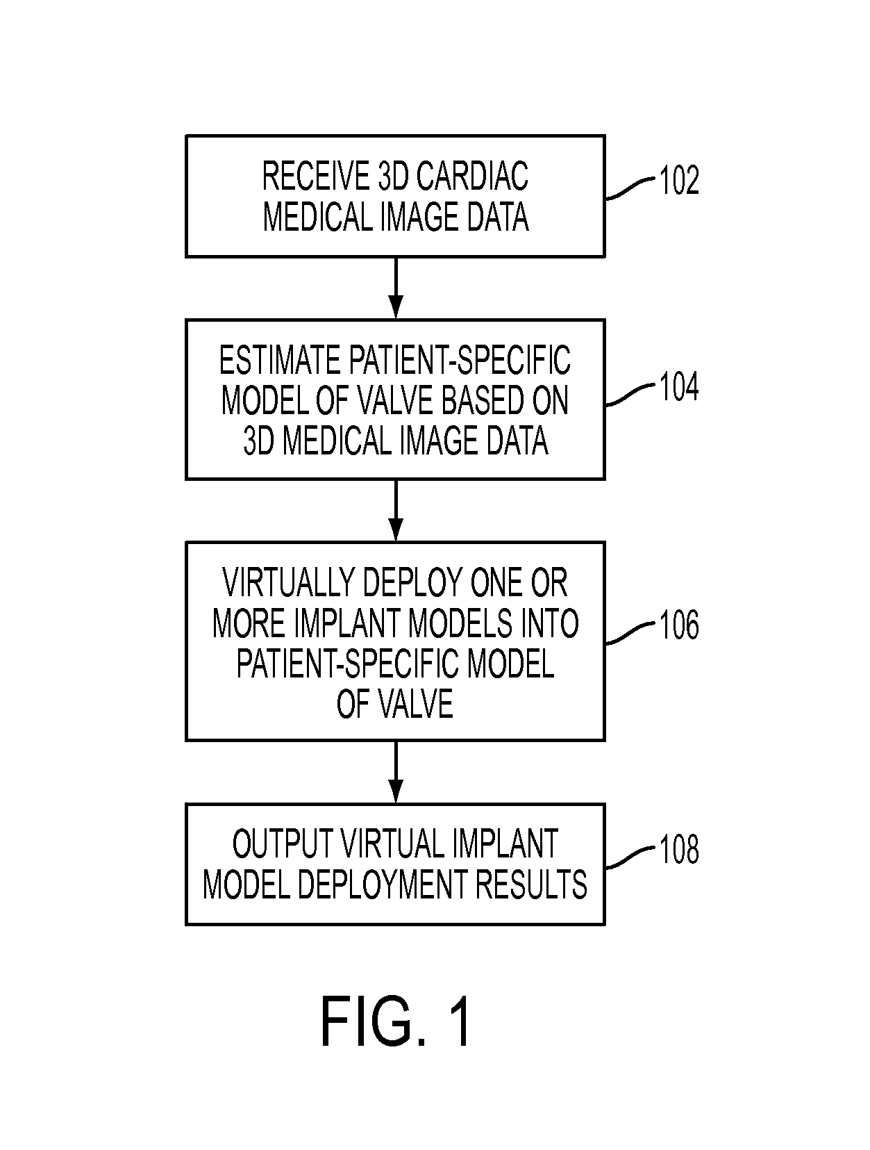
[0005]The present invention provides a method and system of virtual valve implantation for planning, guidance, and assessment of percutaneous valve implantation techniques. Embodiments of the present invention can be used for various types of valve implantation, such as percutaneous aortic valve implantation (PAVI) and percutaneous pulmonary valve implantation (PPVI). Embodiments of the present invention can be used pre-operatively for identification of optimal device type, size and deployment location and for treatment outcome prediction. Embodiments of the present invention can alternatively be used post-operatively quantitative implant assessment.

Problems solved by technology

Traditionally, heart valve replacement has been performed surgically on an open heart, with associated risks including, high mortality, incidence of neurological damage, stroke, and repeated valve replacement.
A misplaced implant can block the coronary ostia and induce a life threatening ischemic condition.
Suboptimal deployment location can result in poor hemodynamic performance with severe paravalvular leakages and / or high gradients and suboptimal effective orifice.
Incorrect implant sizing may require a re-operation and may damage the vessel tissue and cause catastrophic events, such as arterial dissection or rupture.

Method used


Image

  • Method and System for Virtual Percutaneous Valve Implantation
  • Method and System for Virtual Percutaneous Valve Implantation
  • Method and System for Virtual Percutaneous Valve Implantation

Examples


Embodiment Construction

[0017]The present invention relates to a virtual valve implantation framework for modeling and quantitative evaluation of a percutaneous valve implantation procedure. Embodiments of the present invention are described herein to give a visual understanding of the virtual valve implantation method. A digital image is often composed of digital representations of one or more objects (or shapes). The digital representation of an object is often described herein in terms of identifying and manipulating the objects. Such manipulations are virtual manipulations accomplished in the memory or other circuitry / hardware of a computer system. Accordingly, is to be understood that embodiments of the present invention may be performed within a computer system using data stored within the computer system. Embodiments of the present invention are described herein as using 3D or 4D CT data to model patient-specific anatomy. It is to be understood that the present invention is not limited to CT, but ca...


PUM


Abstract

A method and system for virtual percutaneous valve implantation is disclosed. A patient-specific anatomical model of a heart valve is estimated based on 3D cardiac medical image data and an implant model representing a valve implant is virtually deployed into the patient-specific anatomical model of the heart valve. A library of implant models, each modeling geometrical properties of a corresponding valve implant, is maintained. The implant models maintained in the library are virtually deployed into the patient specific anatomical model of the heart valve to select an implant type and size and deployment location and orientation for percutaneous valve implantation.

Description

[0001]This application claims the benefit of U.S. Provisional Application No. 61 / 289,183, filed Dec. 22, 2009, the disclosure of which is herein incorporated by reference.BACKGROUND OF THE INVENTION[0002]The present invention relates to percutaneous valve implantation, and more particularly, to virtual percutaneous valve implantation using medical images.[0003]Valvular heart disease (VHD) is a cardiac disorder that affects a large number of patients and often requires elaborate diagnostic procedures, intervention, and long-term management. Traditionally, heart valve replacement has been performed surgically on an open heart, with associated risks including, high mortality, incidence of neurological damage, stroke, and repeated valve replacement. However, minimally invasive procedures for the valve replacement are less traumatic and reduce the risks associated with valve replacement. Percutaneous valve implantation offers a less invasive alternative to open heart surgery for patients...

Claims


Application Information

Patent Timeline
Patent Type & Authority Applications(United States)
IPC IPC(8): G06F17/50G16H20/40G16H30/20G16H50/50
CPCG06F19/321G06F19/3437G06T2219/2004G06T19/00G06T17/00G16H50/50G16H30/20G16H20/40
Inventor ZAEUNER, DOMINIKIONASEC, RAZVAN IOANGEORGESCU, BOGDANZHENG, YEFENGCOMANICIU, DORINVOIGT, INGMARBOESE, JAN
Owner SIEMENS HEALTHCARE GMBH
Why Eureka
Eureka Blog
Learn More