
Method for assessing the probability of disease development in tissue

Inactive Publication Date: 2011-03-10
MAYO FOUND FOR MEDICAL EDUCATION & RES
View PDF1 Cites 17 Cited by
  • Summary
  • Abstract
  • Description
  • Claims
  • Application Information

AI Technical Summary

Benefits of technology

[0009]The present invention is a method for detecting conditions in tissues that can lead to the development of certain disease states. It is known that abnormal tissue mechanical properties can be a significant cause of certain disease processes, and thus the detection of such abnormal tissue properties can provide a way to predict the development of certain disease states. By detecting such conditions, the present invention enables one to take preemptive actions that terminate or mitigate the conditions, thereby preventing the disease before it develops.

Problems solved by technology

Therefore patients who are found to have such changes in the mechanical properties of otherwise normal tissue may be at much greater risk for eventual development of cancer in those tissues.

Method used


Image

  • Method for assessing the probability of disease development in tissue
  • Method for assessing the probability of disease development in tissue
  • Method for assessing the probability of disease development in tissue

Examples


Embodiment Construction

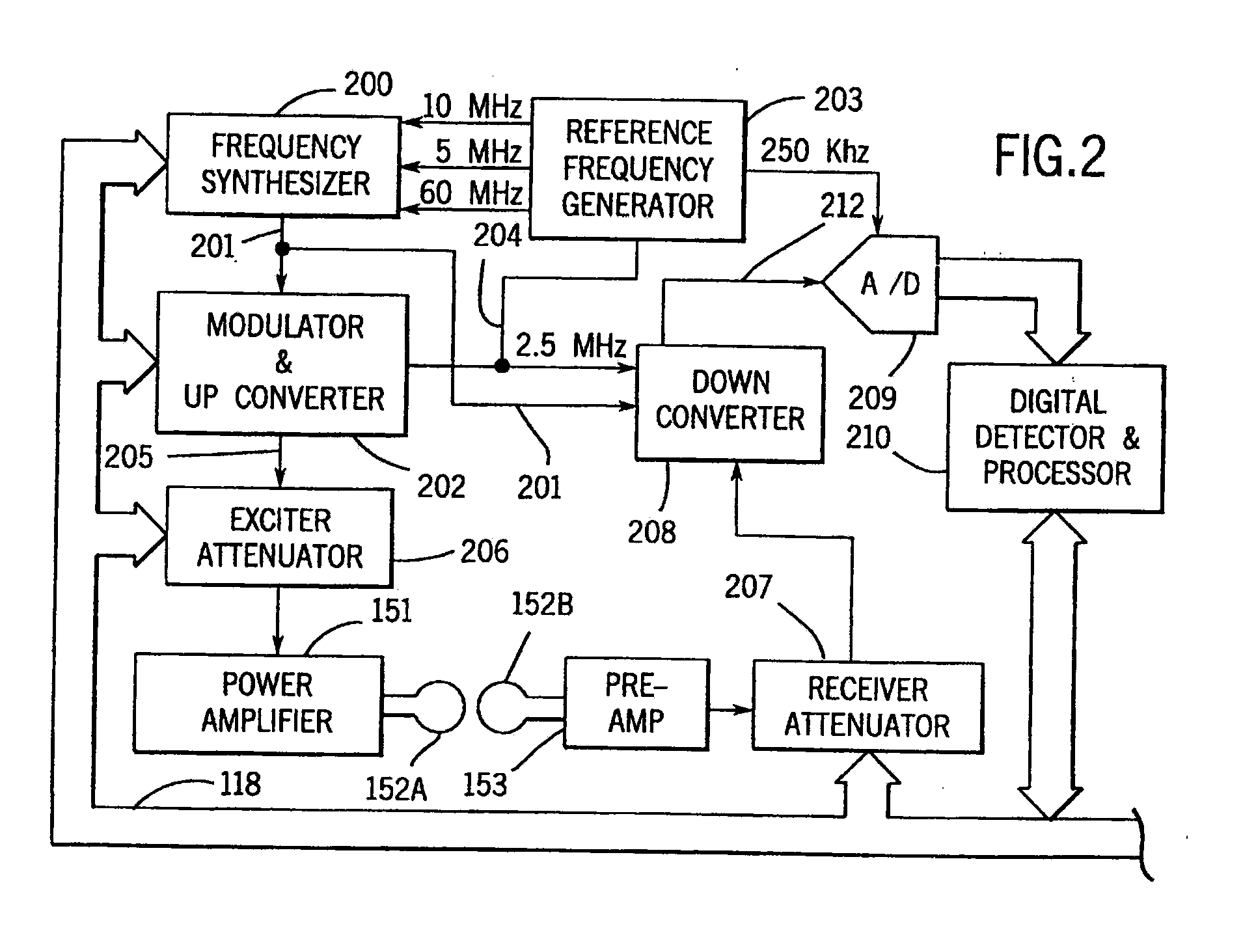
[0019]As indicated above, the present invention may be implemented using any of a number of different techniques for the in vivo measurement of the mechanical characteristics of the tissues of interest. In many cases the choice will be determined by the particular tissues being examined or the particular equipment that is available. Similarly, the particular mechanical characteristic that is measured will be determined not only by the particular tissue being examined, but by the particular disease process of interest. In the preferred embodiment an MRE technique is used to detect the stiffness in organ tissues as a means for predicting the possible onset of fibrosis.

[0020]Referring first to FIG. 1, there is shown the major components of a preferred NMR system which incorporates the present invention and which is sold by the General Electric Company under the trademark “SIGNA”. The operation of the system is controlled from an operator console 100 which includes a console processor 1...


PUM


Abstract

A method for detecting conditions in tissues that can lead to disease includes an in vivo measurement of the mechanical characteristics of the tissues using Magnetic Resonance Elastography (MRE). The deviation of mechanical characteristics such as tissue stiffness from a predetermined norm is determined and indicated on an image of the tissues.

Description

BACKGROUND OF THE INVENTION[0001]The physician has many diagnostic tools at his or her disposal which enable detection and localization of diseased tissues. These include x-ray systems that measure and produce images indicative of the x-ray attenuation of the tissues and ultrasound systems that detect and produce images indicative of tissue echogenicity and the boundaries between structures of differing acoustic properties. Nuclear medicine produces images indicative of those tissues which absorb tracers injected into the patient, as do PET scanners and SPECT scanners. And finally, magnetic resonance imaging (“MRI”) systems produce images indicative of the magnetic properties of tissues. It is fortuitous that many diseased tissues are detected by the physical properties measured by these imaging modalities, but it should not be surprising that many diseases go undetected.[0002]Historically, one of the physician's most valuable diagnostic tools is palpation. By palpating the patient ...

Claims


Application Information

Patent Timeline
IPC IPC(8): A61B5/055A61B5/00
CPCA61B5/0048G01R33/56358A61B5/4244A61B5/055A61B5/0051
Inventor EHMAN, RICHARD L.
Owner MAYO FOUND FOR MEDICAL EDUCATION & RES
Learn More