Method and System for Automatic Contrast Phase Classification

a technology of automatic classification and contrast phase, applied in the field of medical imaging of patients, can solve problems such as insufficient structure or standardization of information

Inactive Publication Date: 2011-01-06
SIEMENS AG
View PDF9 Cites 24 Cited by
  • Summary
  • Abstract
  • Description
  • Claims
  • Application Information

AI Technical Summary

Problems solved by technology

However, the injection of the contrast agent is not typically tied to the image acquisition device used to obtain the medical images.
However, this information is not structured or standardized, and is usually only understandable by a human reader.

Method used


Image

  • Method and System for Automatic Contrast Phase Classification
  • Method and System for Automatic Contrast Phase Classification
  • Method and System for Automatic Contrast Phase Classification

Examples


Embodiment Construction

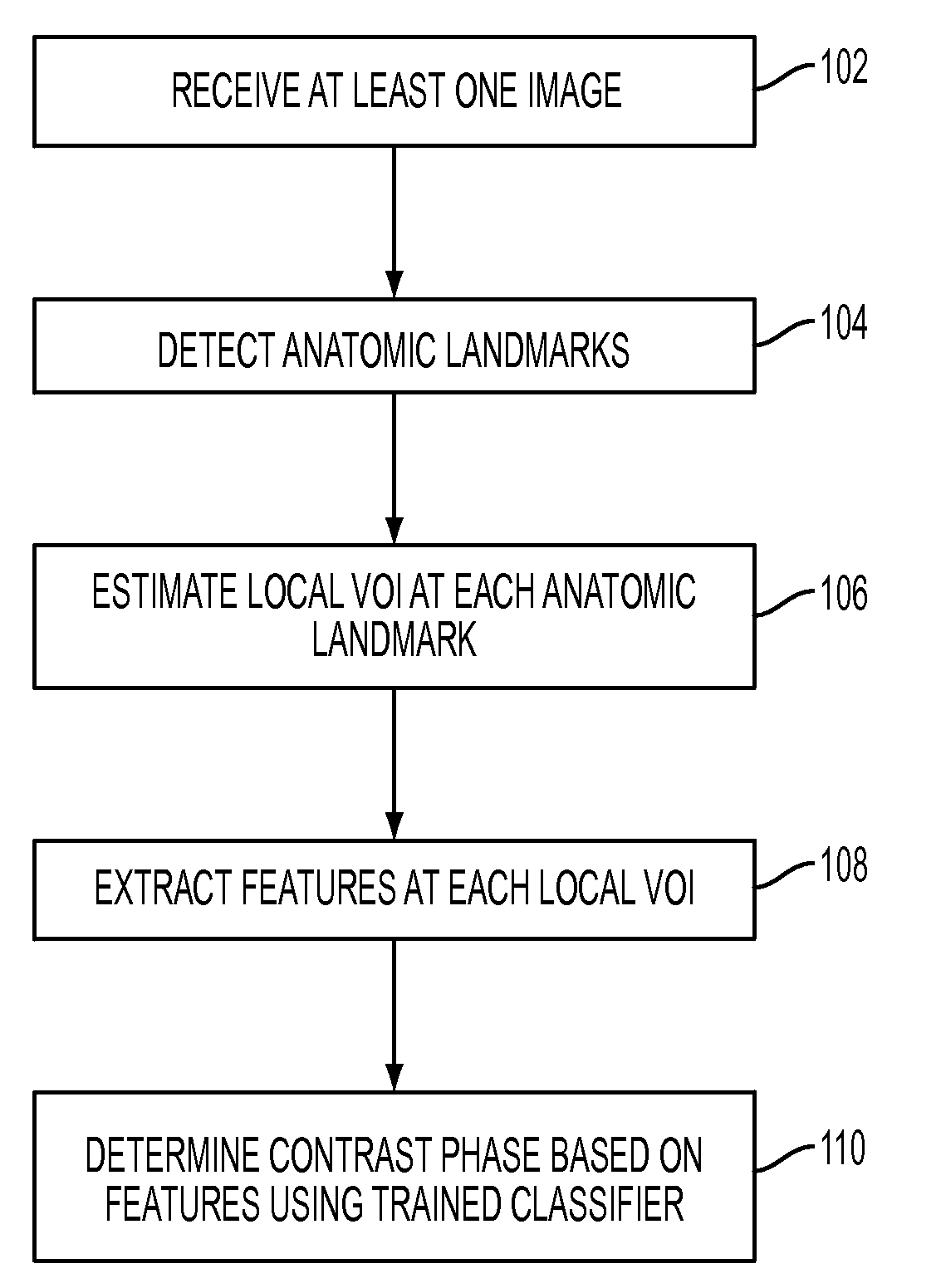
[0016]The present invention is directed to a method and system for automatic classification of a contrast phase in medical images, such as computed tomography (CT) and magnetic resonance (MR) images. As used herein, the “contrast phase” of an image is an indication of when the image was acquired relative to a contrast injection. Embodiments of the present invention are described herein to give a visual understanding of the anatomic landmark detection method. A digital image is often composed of digital representations of one or more objects (or shapes). The digital representation of an object is often described herein in terms of identifying and manipulating the objects. Such manipulations are virtual manipulations accomplished in the memory or other circuitry / hardware of a computer system. Accordingly, it is to be understood that embodiments of the present invention may be performed within a computer system using data stored within the computer system.

[0017]FIG. 1 illustrates a met...


PUM


Abstract

A method and system for classifying a contrast phase of a 3D medical image, such as a computed tomography (CT) image or a magnetic resonance (MR) image, is disclosed. A plurality of anatomic landmarks are detected in a 3D medical image. A local volume of interest is estimated at each of the plurality of anatomic landmarks, and features are extracted from each local volume of interest. The contrast phase of the 3D volume is determined based on the extracted features using a trained classifier.

Description

[0001]This application claims the benefit of U.S. Provisional Application No. 61 / 222,254, filed Jul. 1, 2009, the disclosure of which is herein incorporated by reference.BACKGROUND OF THE INVENTION[0002]The present invention relates to medical imaging of a patient, and more particularly, to automatic classification of a contrast phase in computed tomography (CT) and magnetic resonance (MR) images.[0003]In order to enhance the visibility of various anatomic structures and blood vessels in medical images, a contrast agent is often injected into a patient. Medical images of the patient can be obtained using various imaging modalities, such as CT or MR. However, the injection of the contrast agent is not typically tied to the image acquisition device used to obtain the medical images. Accordingly, medical images typically do not contain contrast phase information regarding how long the image acquisition time was after the contrast injection time.[0004]In clinical routines, contrast phas...

Claims


Application Information

Patent Timeline
IPC IPC(8): G06T7/00
CPCG06T7/0012G06T2207/10081G06T2207/30101G06T2207/30004G06T2207/30008G06T2207/10088
Inventor SUEHLING, MICHAELLIU, DAVIDSOZA, GRZEGORZ
Owner SIEMENS AG
Why Eureka
Try Eureka