Polymer vesicles for selective electromagnetic energy-induced delivery
a technology of electromagnetic energy and polymer vesicles, which is applied in the direction of powder delivery, emulsion delivery, medical preparations, etc., can solve the problems of inability to accurately target the target tissue, damage the surrounding tissue, and inability to focus the microwav
- Summary
- Abstract
- Description
- Claims
- Application Information
AI Technical Summary
Benefits of technology
Problems solved by technology
Method used
Image
Examples
example 1
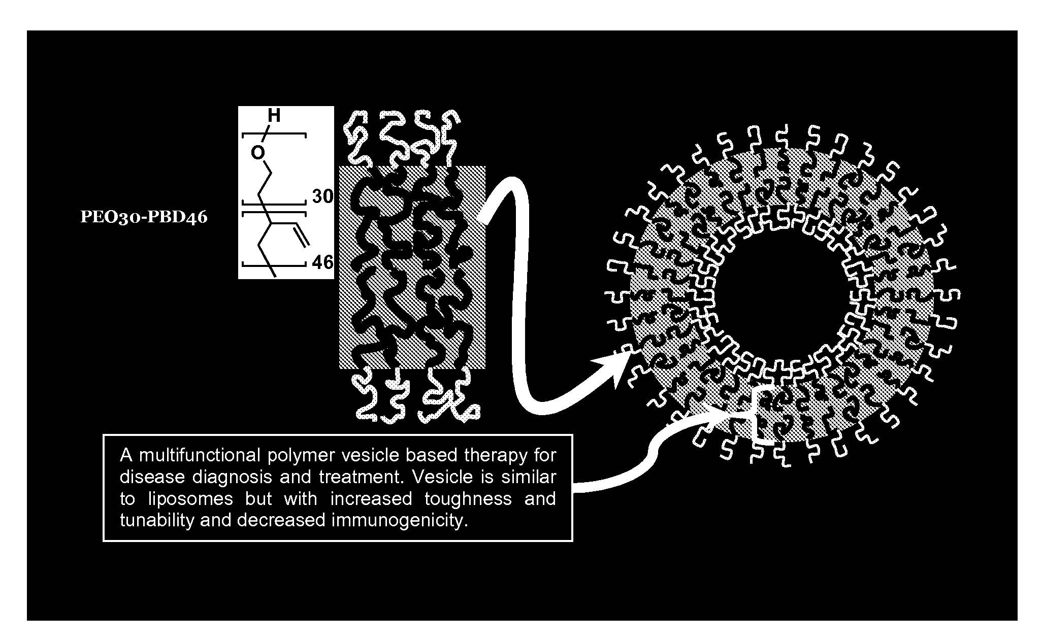
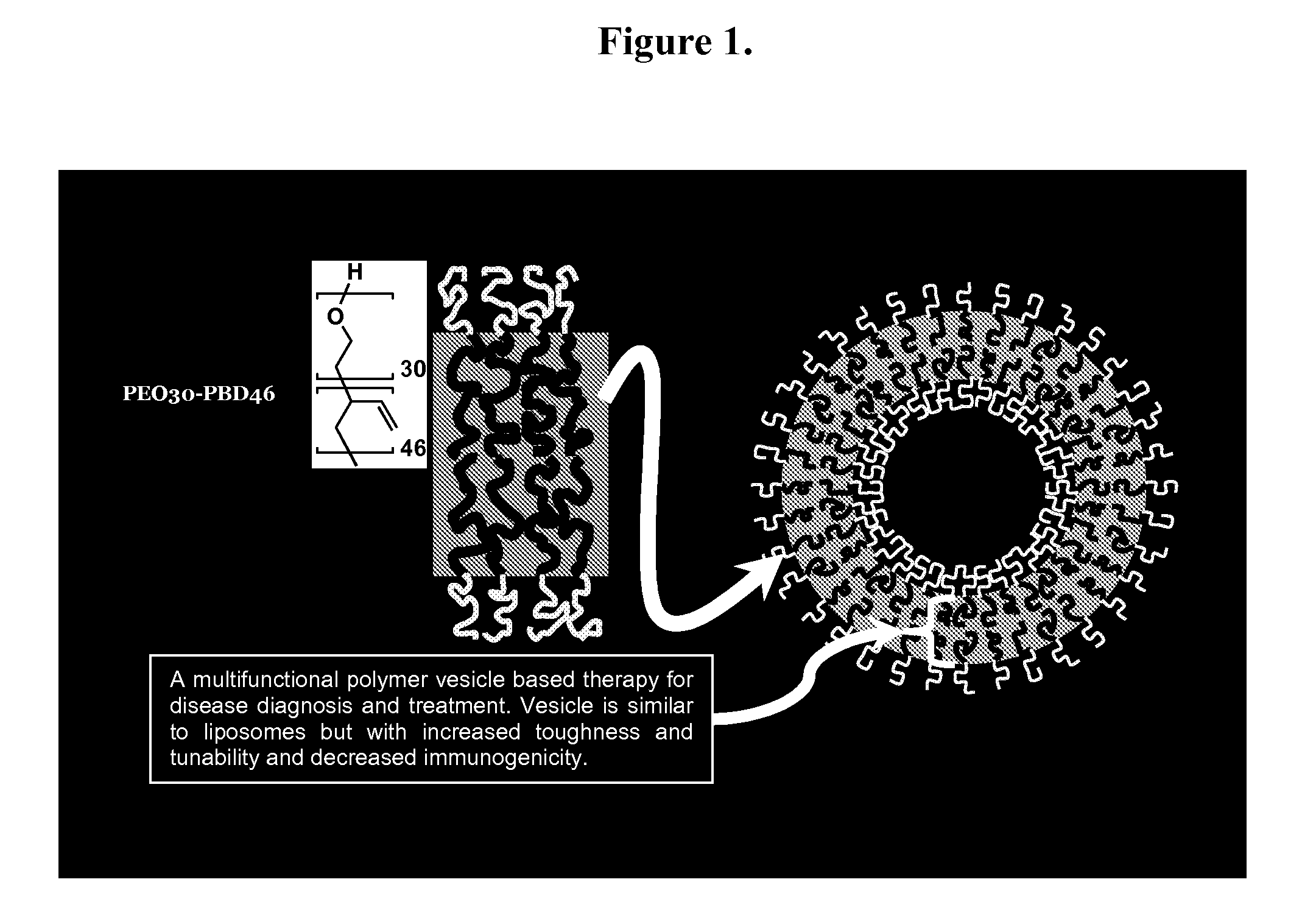
[0073]Polymersome Assembly. Thin films of polymer were created on roughened Teflon surfaces by depositing 200 μL of a 1 mM solution of OB29 with or without porphyrin dye at a 5:1 molar ratio (polymer:dye) in methylene chloride. Films were dried in a vacuum oven for 12 h and then hydrated with 1 or 2 mL of a 290 mOsm sucrose+PBS buffer with or without protein at 65° C. for 24 h. Vesicle solutions were vortexed after heating. Vesicles are depicted in FIG. 1.
example 2
[0074]Vesicle Separation. Vesicles were separated from free protein by diluting 500 μL polymer vesicle solution in 9.5 mL PBS+1% BSA. This solution was then placed on a 2 mL cushion of sucrose buffer+density gradient medium in a centrifuge tube. The tube was spun at 20,000 rpm for 1 h, and the resulting band of 500-1000 μL concentrated solution of vesicles was removed. Samples were then dialyzed into 290 mOsm PBS within 20,000 Da MWCO dialysis cassettes for 12 h at 4° C.
example 3
[0075]Chemical labeling of HSF, HSAF, BSA and Mb. The concentration of HSAF, BSA, or Mb was determined by UV-Vis spectroscopy (ε280=480,000 M−1cm−1 for HSAF,[2]ε280=44,300 M−1cm−1 for BSA, and £280=15,400 M−1cm−1 for Mb). BSA required several FPLC size-exclusion purification steps to eliminate protein aggregates and fragments. The concentration of HSF was determined by Bradford assay. The Cy3 dye was used to label surface lysines on the protein, whereas BODIPY-FL labeled surface cysteines. The protein labeling protocols provided by the manufacturers were followed for both Cy3 and BODIPY-FL conjugations, with the following modifications. For the Cy3 labeling: i) 10 mg / mL protein in 290 mOsm PBS was used, ii) the conjugation reaction was carried out for 2-3 h, and iii) the labeled protein was separated from free dye with 290 mOsm sucrose+PBS buffer. For the BODIPY-FL labeling: i) 500-fold molar excess (20-fold per ferritin subunit) of TCEP was used, ii) 400-fold molar excess (17-fold ...
PUM
 Abstract
Description
Claims
Application Information
 - R&D
- Intellectual Property
- Life Sciences
- Materials
- Tech Scout
- Unparalleled Data Quality
- Higher Quality Content
- 60% Fewer Hallucinations
Browse by: Latest US Patents, China's latest patents, Technical Efficacy Thesaurus, Application Domain, Technology Topic, Popular Technical Reports.
© 2025 PatSnap. All rights reserved.Legal|Privacy policy|Modern Slavery Act Transparency Statement|Sitemap|About US| Contact US: help@patsnap.com


