
Diagnostic ultrasound apparatus

a diagnostic ultrasound and ultrasound technology, applied in diagnostics, medical science, applications, etc., can solve problems such as limitations in terms

Inactive Publication Date: 2009-12-03
CANON KK
View PDF8 Cites 29 Cited by
  • Summary
  • Abstract
  • Description
  • Claims
  • Application Information

AI Technical Summary

Benefits of technology

[0008]However, with a diagnostic ultrasound apparatus described in Japanese Patent Application Laid-Open No. 2005-137581, information relating to the probe positions obtained from the sensors attached to the respective probes is employed only when synthetically combining the images obtained from the probes substantially independently. The probes operate only for focusing from the predetermined respective imaging angles of view. In other words, the shooting operation of each o...

Problems solved by technology

Therefore, as disclosed in the patent document, even if the plurality of probes are arranged flexibly to accommodate themselves to the profile of the body surface, they are subjected to limitations in terms of directions and angles that should be selected to obtain cross-sectional images that are effective for diagnosing the desired living tissue.

Method used


Image

  • Diagnostic ultrasound apparatus
  • Diagnostic ultrasound apparatus
  • Diagnostic ultrasound apparatus

Examples


example

[0142]Now, an example where the present invention is applied to blood flow measurement of a diagnostic ultrasound apparatus will be described below by referring to FIG. 4.

[0143]The arrangement of this example can be used for a measurement operation of determining the moving speed of tissue or the flowing speed of blood in a blood vessel at a specific position that is selected as the moving part of a specific body portion.

[0144]While an operation of measuring the blood flow rate is described below as a typical example, the following description is generally applicable to any operation of measuring the speed of a moving part of a body. The Doppler method or the color Doppler method is employed in conventional diagnostic ultrasound apparatus to measure the blood flow rate and determine the blood flow volume by using the measured blood flow rate. However, since the Doppler method and the color Doppler method are methods of detecting the phase difference of ultrasonic echo signals, they ...


PUM


Abstract

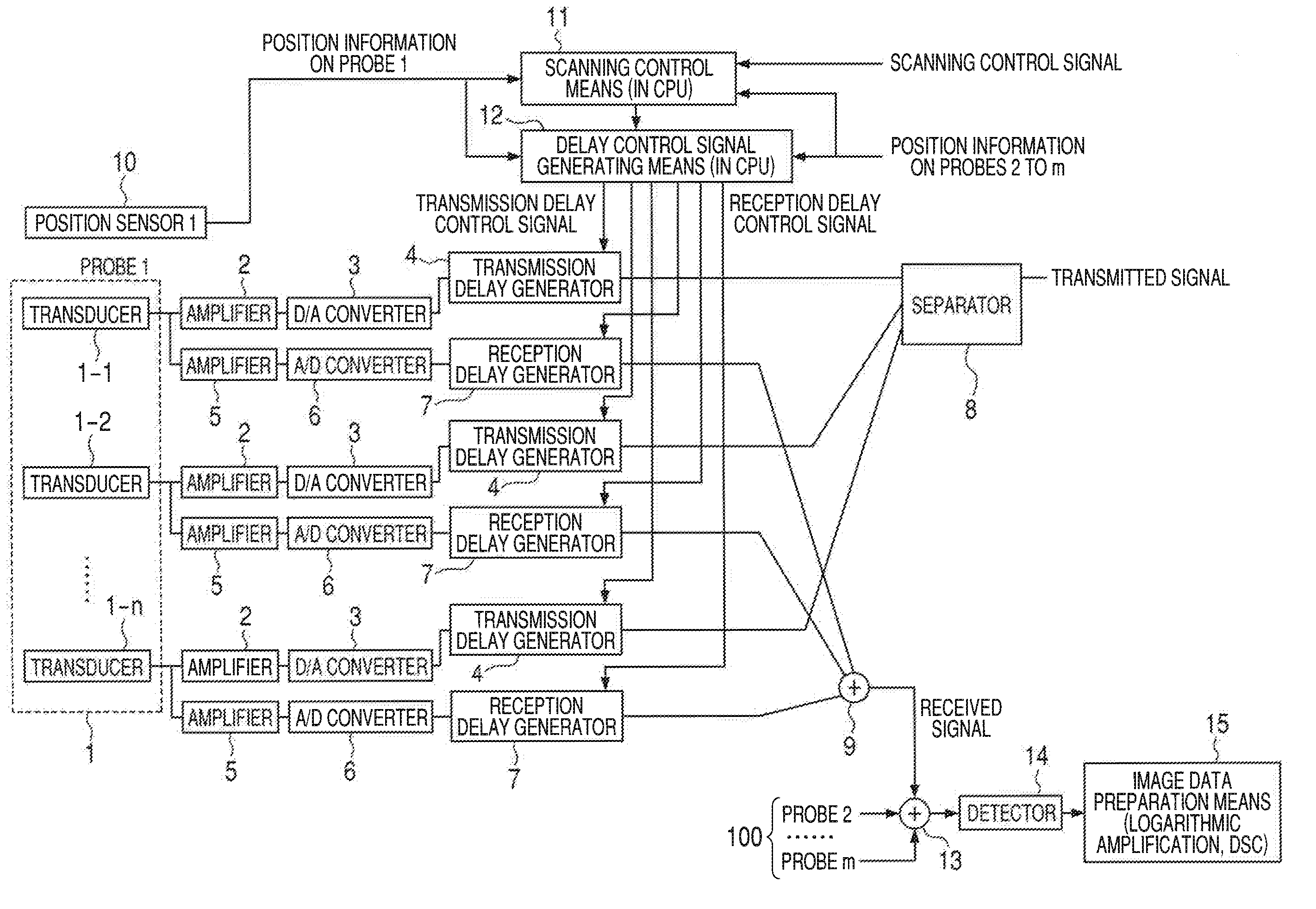
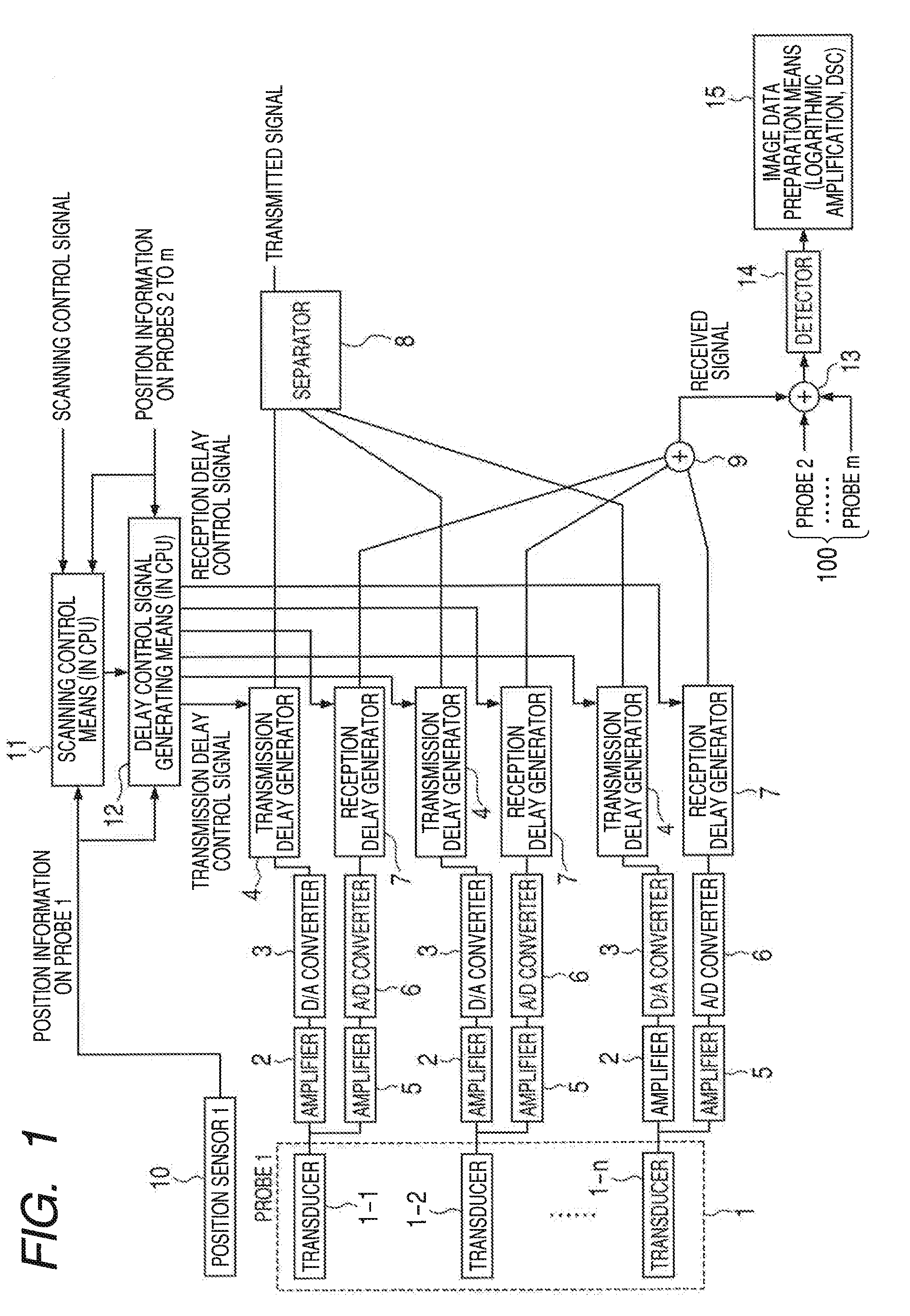
A novel diagnostic ultrasound apparatus has a plurality of ultrasound probes 1, delay means pairs 4 and 7 arranged respectively in the plurality of ultrasound probes to electronically delay the timings of transmissions or receptions of ultrasonic waves by the ultrasonic transducers, position / orientation detecting means 10 and delay control signal generating means for generating a delay control signal to control signal delays of the plurality of ultrasonic transducers of the plurality of ultrasound probes, using information acquired from the means 10. The delay control signal is input to the delay means to control the timings of transmission of ultrasonic waves to a target object of examination.

Description

BACKGROUND OF THE INVENTION[0001]1. Field of the Invention[0002]This invention relates to a diagnostic ultrasound apparatus and a diagnostic imaging apparatus for medical use designed to prepare image data according to an ultrasonic echo signal.[0003]2. Description of the Related Art[0004]Diagnostic ultrasound apparatus for transmitting ultrasonic waves from a plurality of probes, acquiring ultrasonic echo signals and synthesizing a cross-sectional image of living tissue so as to collect many pieces of information more easily than an apparatus using a single probe are known.[0005]Japanese Patent Application Laid-Open No. 2005-137581 discloses a moving image photographing apparatus for acquiring a cross-sectional image of living tissue that has a plurality of ultrasound probes, anchors for rigidly securing them, an acoustic coupling material, a probe switching section, an image data measuring section, an image data synthesizing section and a moving image displaying section.[0006]Each...

Claims


Application Information

Patent Timeline
IPC IPC(8): A61B8/14
CPCA61B8/4254A61B8/14A61B8/4209A61B8/4477A61B8/54
Inventor OIKAWA, KATSUYANAGAE, KENICHISOMEDA, YASUHIRO
Owner CANON KK
Learn More