
Two-dimensional or three-dimensional imaging of a target region in a hollow organ

a target region technology, applied in the field of two-dimensional or three-dimensional imaging of a target region in a hollow organ, can solve the problems of difficult to achieve, take a very long time,

Inactive Publication Date: 2008-07-24
SIEMENS AG
View PDF4 Cites 98 Cited by
  • Summary
  • Abstract
  • Description
  • Claims
  • Application Information

AI Technical Summary

Benefits of technology

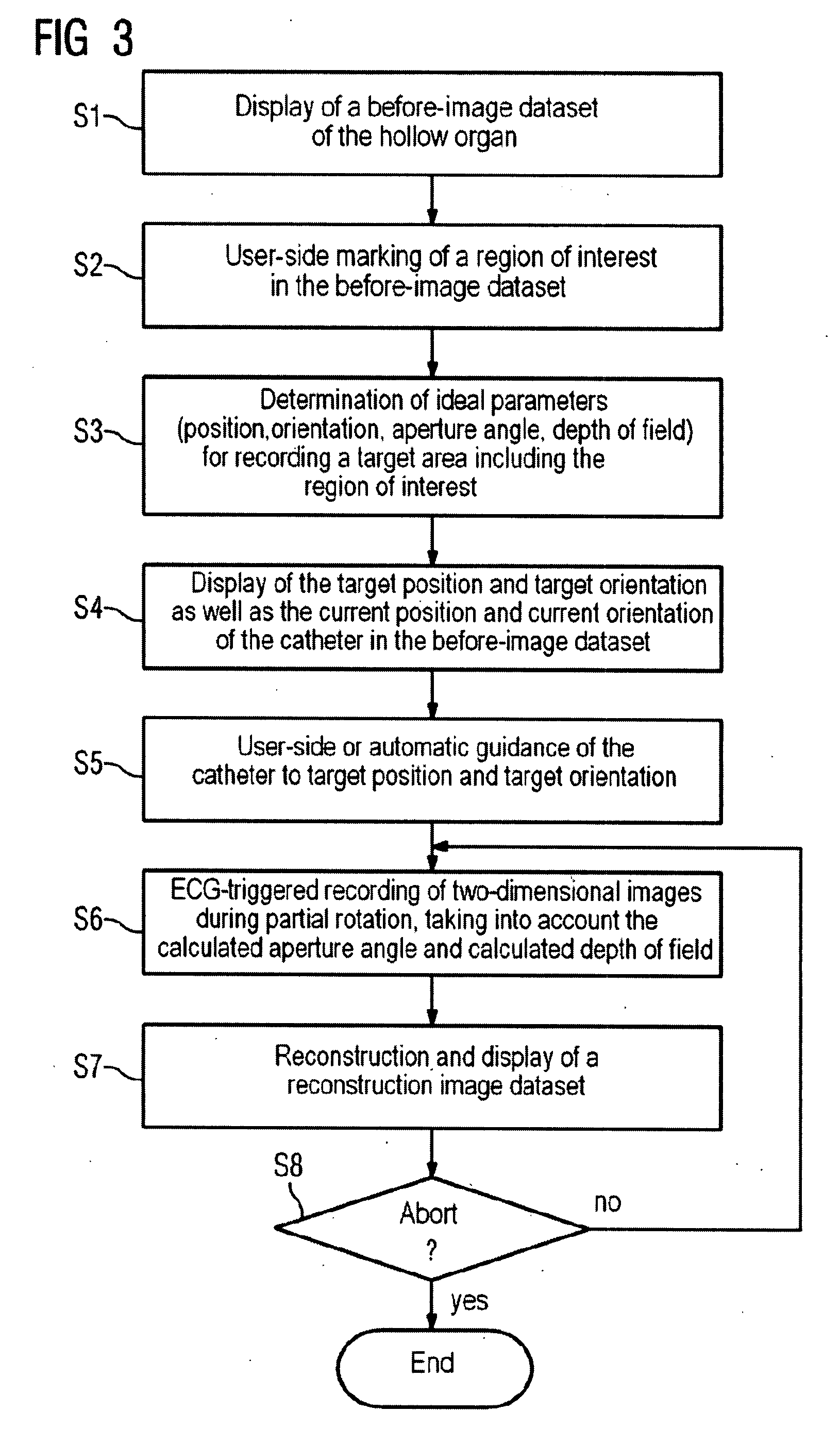
"The invention provides a method for faster and more efficient reconstruction and visualization of a target region of interest in a hollow organ, such as the heart, by recording images during a partial rotation of the image recording device through a specific angle. This approach reduces the number of images needed and allows for more frequent updates of the reconstruction image dataset. The method can be used in real-time interventional procedures and can provide a faster and more accurate representation of the target region."

Problems solved by technology

However, difficulties arise here if the images are to be displayed in realtime, in particular in the case of rhythmically moving hollow organs, in particular in the region of the heart, where only two-dimensional single images recorded within the same phase in different ECG cycles can be used for the reconstruction image dataset.
The recording of the two-dimensional single images during a full rotation of the image recording device through 360° therefore takes a very long time which can extend for example over several ECG cycles.
A further disadvantage arises from the fact that in addition to the target region of interest, as a result of the recording of the complete hollow organ a multiplicity of image information is recorded which ultimately is irrelevant to the actual medical problem under consideration.

Method used


Image

  • Two-dimensional or three-dimensional imaging of a target region in a hollow organ
  • Two-dimensional or three-dimensional imaging of a target region in a hollow organ
  • Two-dimensional or three-dimensional imaging of a target region in a hollow organ

Examples


Embodiment Construction

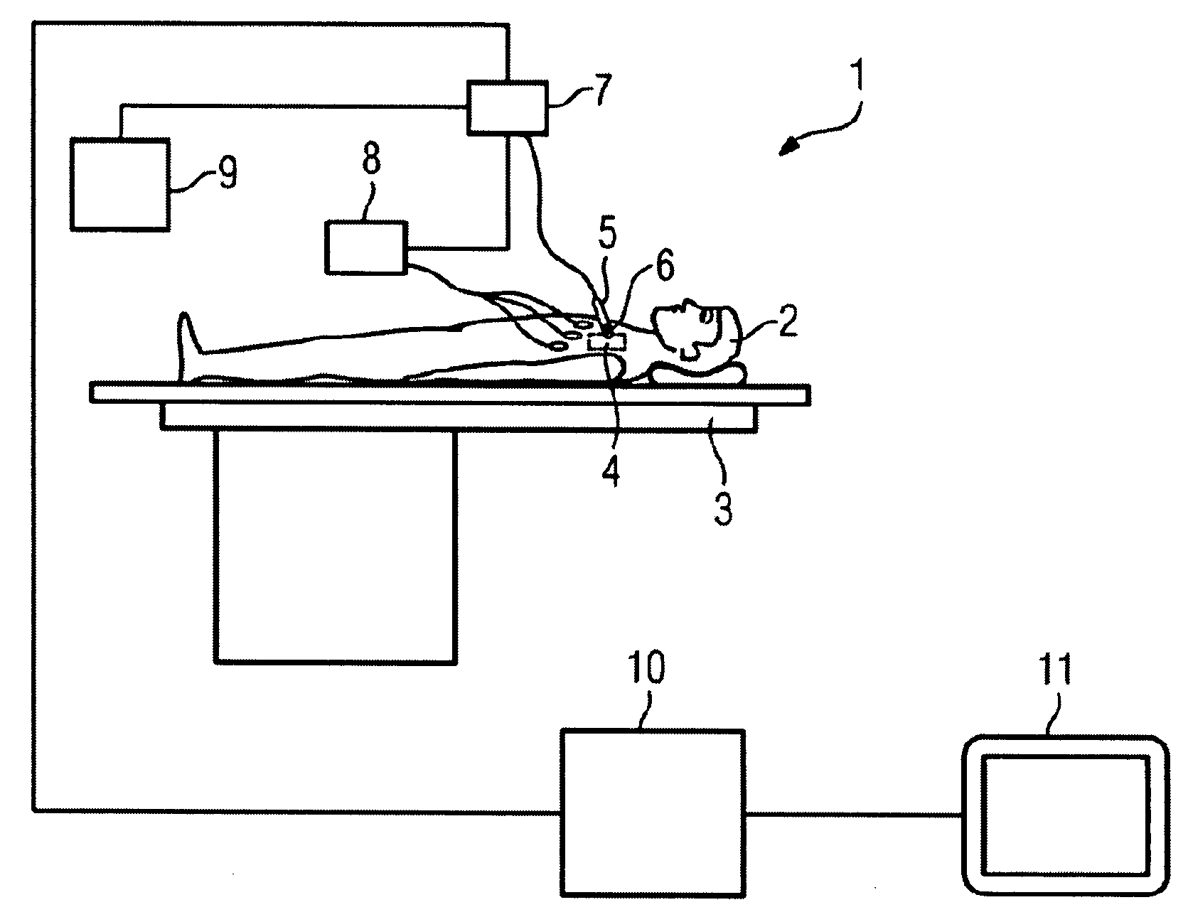
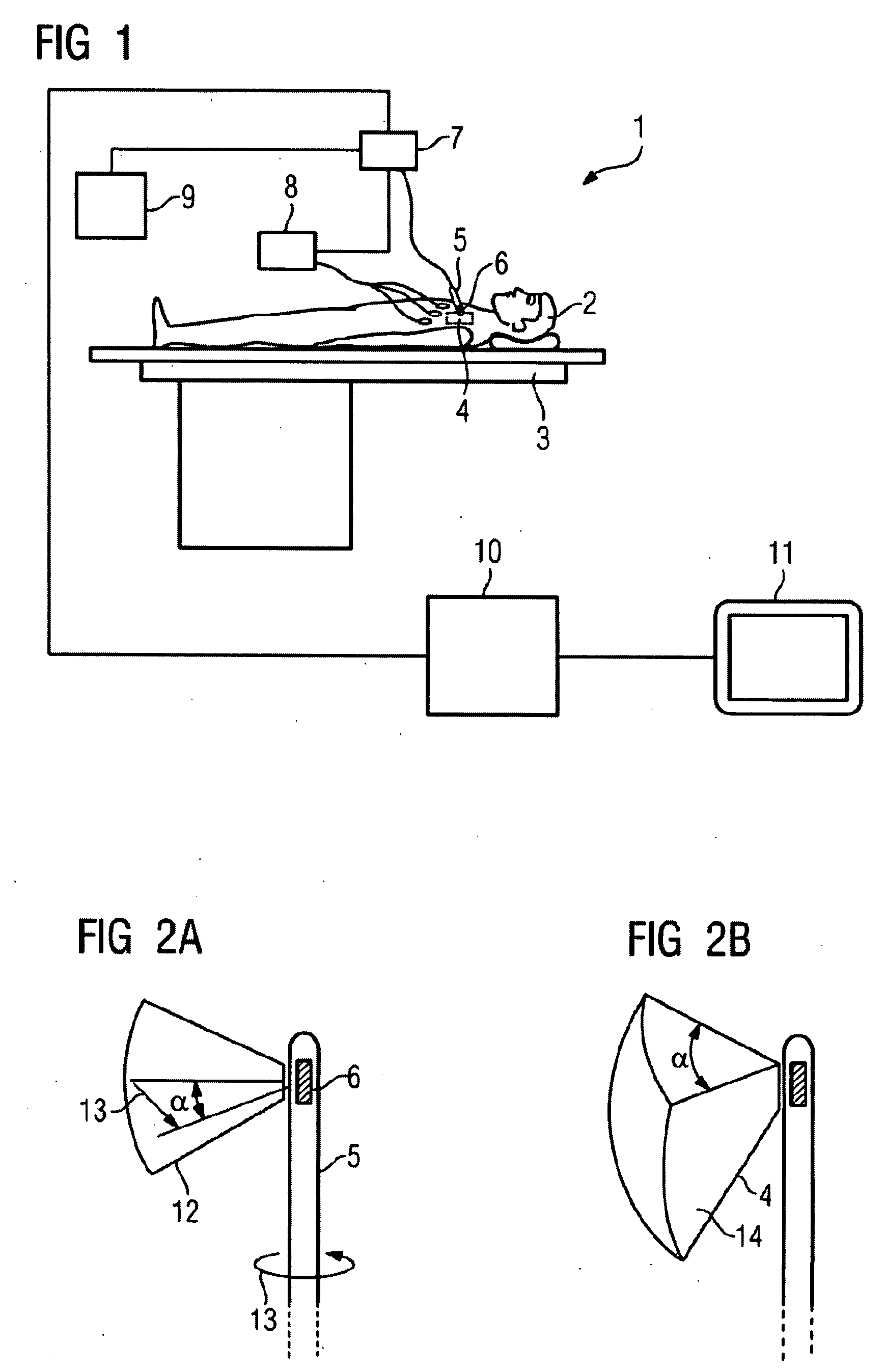
[0045]FIG. 1 shows a medical examination or treatment system 1. In this example a patient 2 is lying on a positioning table 3, the patient including a target region 4 that is to be recorded, in this case in particular in the cardial region, into which a catheter 5, comprising a rotatably disposed image recording device 6, has been introduced. The catheter 5 and the image recording device 6 are controlled accordingly by means of a catheter control device 7, though a guiding of the catheter by hand is also possible. An ECG device 8 records the ECG of the patient 2. The ECG device 8 sends its data to the catheter control device 7 so that a triggered recording of two-dimensional images by means of the image recording device 6, in this example an ultrasound device, can be performed. The position and orientation of the image recording device of the catheter 5 can be determined at any time by means of a navigation system indicated by the reference numeral 9. The control device 7 communicat...


PUM


Abstract

A two-dimensional or three-dimensional imaging of a target region in a hollow organ is provided. A two- or three-dimensional reconstruction image dataset is reconstructed from two-dimensional images from the inside of the hollow organ that are recorded by via a rotating image recording device and displayed, with images covering the entire target region being recorded during a partial rotation of the image recording device through a rotation angle.

Description

CROSS REFERENCE TO RELATED APPLICATIONS[0001]This application claims priority of German application No. 102006046045.6 DE filed Sep. 28, 2006, which is incorporated by reference herein in its entirety.FIELD OF INVENTION[0002]The invention relates to a method for the two-dimensional or three-dimensional imaging of a target region in a hollow organ, wherein a two- or three-dimensional reconstruction image dataset is reconstructed from two-dimensional images from the inside of the hollow organ that are recorded by means of a rotating image recording device and displayed.BACKGROUND OF INVENTION[0003]In examinations or minimally invasive interventions in hollow organs, in particular in the region of the heart, medical instruments are used which include an image recording device. By means of the images of said image recording device it is aimed to examine a target region of a hollow organ or to monitor an intervention that is taking place therein. Since it has been customary in the past t...

Claims


Application Information

Patent Timeline
Patent Type & Authority Applications(United States)
IPC IPC(8): A61B5/055
CPCA61B19/52A61B2017/00044A61B2017/00243A61B2019/501A61B2019/502A61B2019/5295A61B2019/5251A61B2019/5276A61B2019/528A61B2019/5289A61B2019/5291A61B2019/507A61B2034/101A61B2034/102A61B90/36A61B2090/367A61B2090/364A61B2090/3784A61B2034/107A61B2034/2051A61B2090/378A61B2090/365
Inventor JOHN, MATTHIASRAHN, NORBERT
Owner SIEMENS AG
Learn More