
Enhanced navigational tools for comparing medical images

a technology of navigational tools and medical images, applied in image enhancement, medical/anatomical pattern recognition, instruments, etc., can solve the problems of difficult to find and match the corresponding nodules in the stack of hundreds of axial slices, and the process of tracking and comparing nodules across multiple temporal ct scans is very tedious and time-consuming, so as to facilitate the matching and comparative visualization process and achieve different visual appearance

Active Publication Date: 2007-08-02
MEVIS MEDICAL SOLUTIONS
View PDF4 Cites 55 Cited by
  • Summary
  • Abstract
  • Description
  • Claims
  • Application Information

AI Technical Summary

Benefits of technology

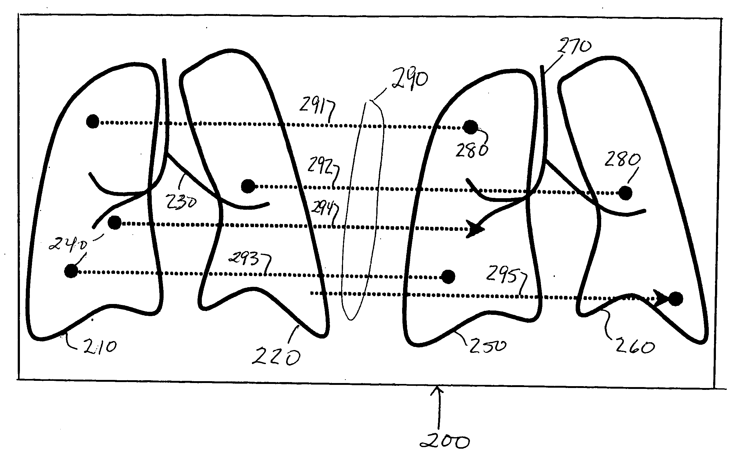
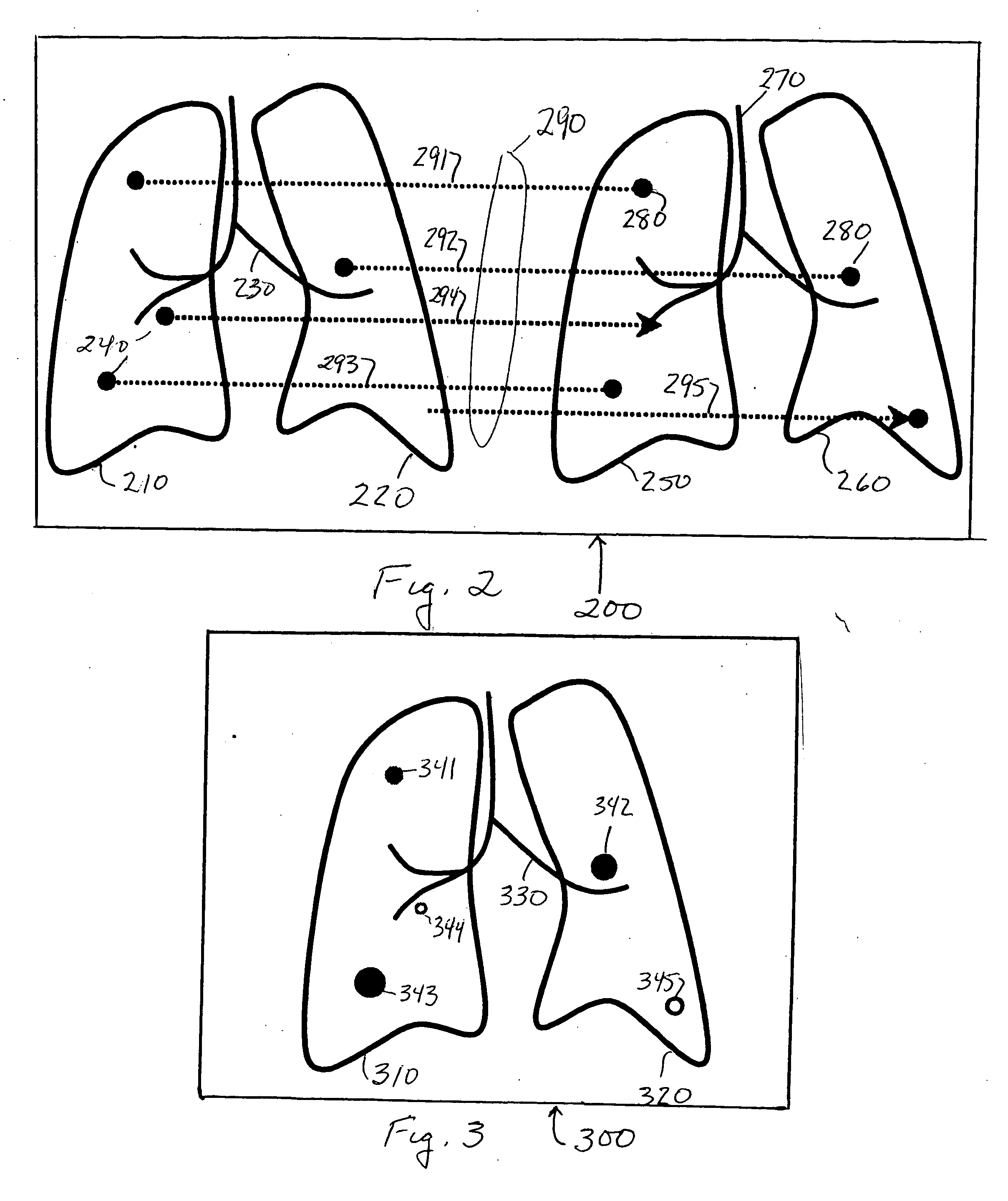
[0004] The present invention is directed to a navigation tool that facilitates this matching and comparative visualization process. In an illustrative embodiment of the invention, the tool provides a side-by-side computer-driven display of two temporally related medical images of the same region of a patient. A plurality of lines extend between the images, at least a first line beginning at a feature at a first location in a first image and ending at a corresponding feature at a corresponding location in the second image and at least a seco...

Problems solved by technology

In today's clinical practice, the tracking and comparing of nodules across multiple temporal CT scans is a very tedious process.
While numerous tools exist for matching the major anatomical features of the lungs that are visible in both stacks, it is extremely time-consuming to find and match the corresponding nodules in the two stacks of hundreds of axial slices.

Method used


Image

  • Enhanced navigational tools for comparing medical images
  • Enhanced navigational tools for comparing medical images
  • Enhanced navigational tools for comparing medical images

Examples


Embodiment Construction

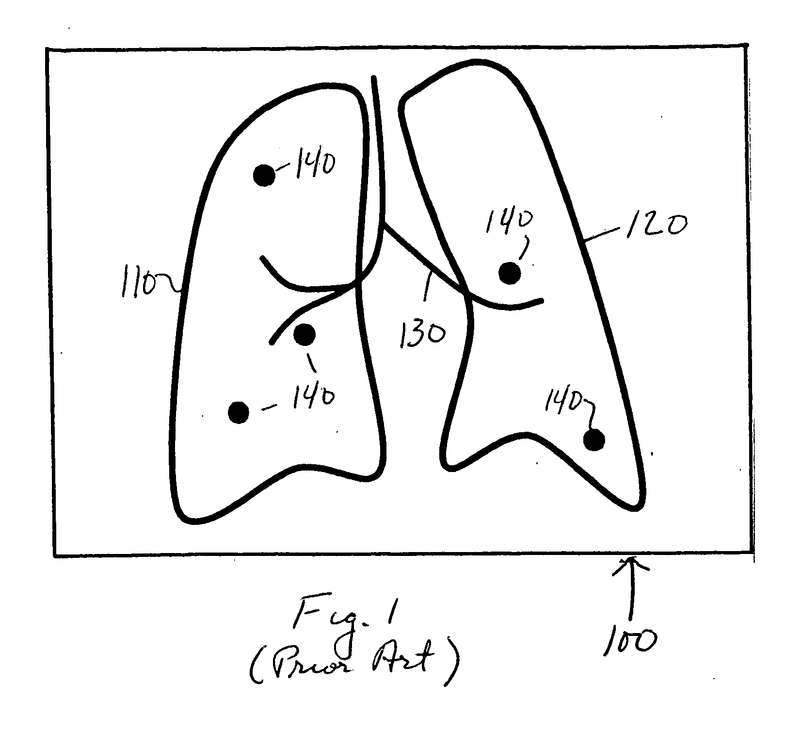
[0014]FIG. 1 depicts a display 100 of nodules on a lung anatomical background map constructed to facilitate localization and comparative visualization of the nodules. Using sophisticated image processing and segmentation tools, key anatomical structures can be extracted from the original x-rays, CT axial sections and the like and represented in a map in several ways. For example, one can project the anatomical structures onto a 2D plane and create a projection 2D map. Another method, as shown here, is to create a line drawing type of map to represent the lung anatomy. The border of the lungs is represented by closed lines 110, 120. Other anatomical background such as the airway and vascular structures are represented by dark lines 130. The approximate location of nodules relative to the background structures represented by lines 110, 120, 130 is shown by discs 140. Advantageously, the discs are brightly colored, e.g., red, to make them stand out in the display. Alternatively, the di...


PUM


Abstract

A navigation tool that provides a side-by-side display of two temporally related medical images of the same region of a patient. A plurality of lines extend between the images, at least a first line beginning at a feature at a first location in a first image and ending at a corresponding feature at a corresponding location in the second image and at least a second line extending from a feature found at a second location in one of the images toward a corresponding second location in the other image where no corresponding feature is found. Thus, the second line indicates a feature found in one image but not the other. Advantageously, the first and second lines have different visual appearance. For example, they are different colors. Preferably, the navigational tool also includes a scroll bar alongside the display of medical images. On the scroll bar are a series of markers, each marker corresponding to one of the plurality of lines that extend between the images and preferably being aligned with that line.

Description

BACKGROUND OF THE INVENTION [0001] This relates to navigational tools to facilitate the comparison of two temporally related medical images on a computer-driven display. The term medical images is used broadly herein to refer not only to actual images such as x-rays, CT axial sections, MRI images, sonograms, mammograms and the like, but also to representations and / or abstractions of such images as in the case of a display of regions of interest on an anatomical background map. The invention is particularly useful in the comparison of chest computer tomography (CT) scans of the same patient taken at different times and will be described in that context. The principles of the invention may, however, be applied in the comparison of any two medical images of the same features. [0002] It is common clinical practice to acquire chest CT scans of the same patient at different times and compare them to monitor nodular structures. By comparing the potentially pathological nodular structures p...

Claims


Application Information

Patent Timeline
IPC IPC(8): G06K9/00G16H30/20G16H30/40
CPCG06F19/321G06F19/3406G06K2209/05G06T7/0016G06T2219/028G06T2200/24G06T2207/10072G06T2207/10116G06T2207/30064G06T19/00G16H40/63G16H30/20G16H30/40G06V2201/03
Inventor CHUI, HAILI
Owner MEVIS MEDICAL SOLUTIONS
Learn More