Endoscopic instruments
a technology of endoscopic instruments and valves, applied in the field of endoscopic instruments, can solve the problems of insufficient thickness and large volume of the valve formed in this application, accompanied by serious pain, and insufficient reflux prevention treatment with medication, so as to achieve the effect of preventing gastroesophageal reflux
- Summary
- Abstract
- Description
- Claims
- Application Information
AI Technical Summary
Benefits of technology
Problems solved by technology
Method used
Image
Examples
first embodiment (
FIGS. 1 to 20, 29)
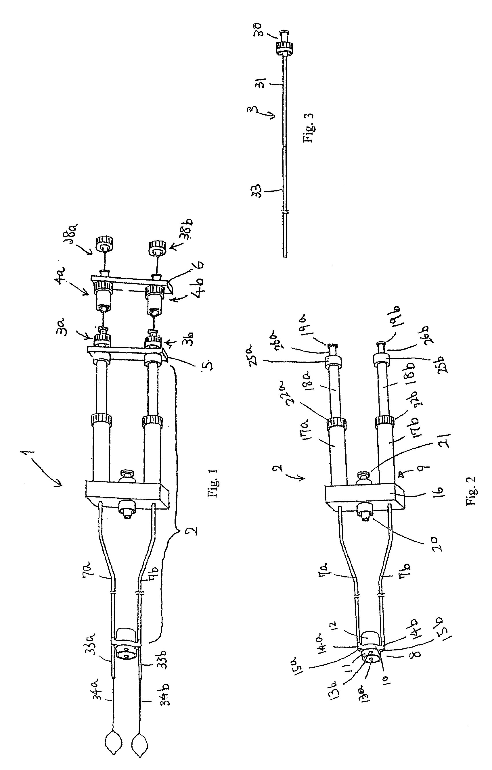
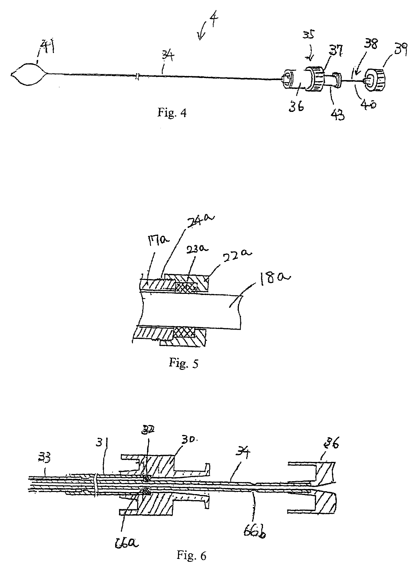
Description of a Piercing Device and a Knot Pusher (FIGS. 1 to 7,10, 29)
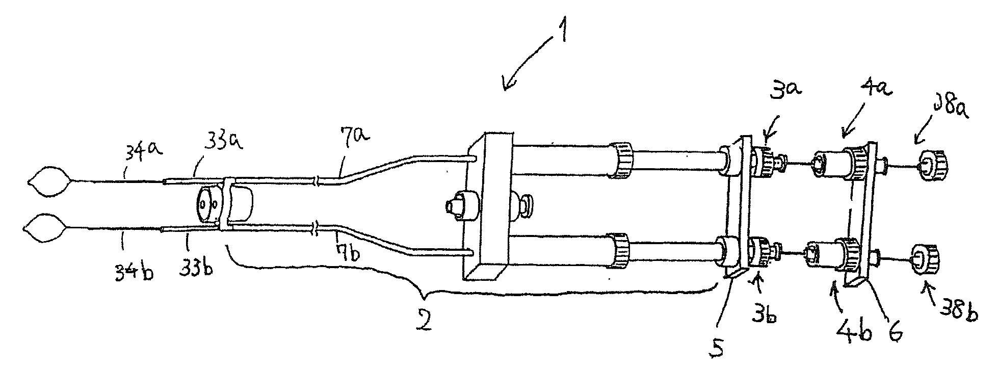
[0057] A piercing device 1 comprises a main body 2, inner sheaths 3a and 3b, needles 4a and 4b, an inner sheaths-coupling member 5, and a needles-coupling member 6. [0058] The main body 2 comprises two outer sheaths 7a and 7b, caps 8 connected at the distal ends of the sheaths 7a and 7b, and an operation section 9 connected at the proximal end of the sheaths 7a and 7b.
[0059] The cap 8 comprises an outer sheaths-connecting section 10, a distal cylindrical section 11, and a distal mounting section 12.
[0060] The distal cylindrical section 11 is made of a relatively hard material. Preferably, it is made of a transparent plastic material such as polycarbonate lest it should obstruct the vision of a first endoscope 27. Preferably, the inner diameter is about 5 to 15 mm, the wall thickness is about 1 mm. The length is about 3 to 10 mm, and a shorter cylinder is better. Side holes 13a and 13b are po...
second embodiment (
FIGS. 21 to 27)
[0143] Only points which are different from those of the first embodiment are described.
[0144] A piercing device 101 is the piercing device 1 with its needles 4a and 4b replaced by needles 102a and 102b.
[0145] The needle 102 comprising a needle body 34, a needle grip 104 connected proximal to the needle body 34, and a suture 103 inserted movably in the needle body 34 and the needle grip 104. The needle grip 104 has a narrow section 43 to which the needles-coupling member 6 is connected.
[0146] The lumen in the outer channel 51 fixed on the outer periphery of the second endoscope 57 has suture-holding forceps 105 which are inserted movably.
[0147] The suture-holding forceps 105 comprise a flexible sheath 107, an operation section 108 which is rotated against and connected proximal to the flexible sheath 107, a handle 109 which slides against the operation section (108), a driving member 110 which is connected at the distal end of the handle 109 and extended slidably ...
third embodiment (
FIGS. 28 and 30)
[0158] Only points which are different from those of the first embodiment are described.
[0159] The suture-holding forceps 121 of the needle 120 have a holding section 122 at the distal end of the operation pipe 40. The holding section 122 is bent and mounted to the operation pipe 40 so that the central axis 123 of the loop 124 forming the holding section 122 inclines against the longitudinal axis of the operation pipe 40.
[0160] When the holding section 122 is rotated, the loop 124 forming the holding section 122 is moved in an area 125 larger than that of the first embodiment.
[0161] Because the loop 124 forming the holding section 122 moves in a larger area, the suture-end is inserted into the loop 124 easily for improved operability and shortened time.
PUM
 Abstract
Description
Claims
Application Information
 - R&D
- Intellectual Property
- Life Sciences
- Materials
- Tech Scout
- Unparalleled Data Quality
- Higher Quality Content
- 60% Fewer Hallucinations
Browse by: Latest US Patents, China's latest patents, Technical Efficacy Thesaurus, Application Domain, Technology Topic, Popular Technical Reports.
© 2025 PatSnap. All rights reserved.Legal|Privacy policy|Modern Slavery Act Transparency Statement|Sitemap|About US| Contact US: help@patsnap.com


