
Laparoscopic port site closure tool

a port site and tool technology, applied in the field of surgical suturing devices, can solve the problems of insufficient closure of surgical defects, difficult and time-consuming incision closure, and herniation of omentum or bowel, and achieve the effect of simple and quick operation

Active Publication Date: 2006-02-09
TELEFLEX MEDICAL INC
View PDF100 Cites 132 Cited by
  • Summary
  • Abstract
  • Description
  • Claims
  • Application Information

AI Technical Summary

Benefits of technology

[0011] In view of these needs, the present invention contemplates a closure tool that in the first instance applies pressure to the suture site from inside the abdominal cavity, and in the second instance provides a simple, quick-operating mechanism for passing the suture through the body tissue.
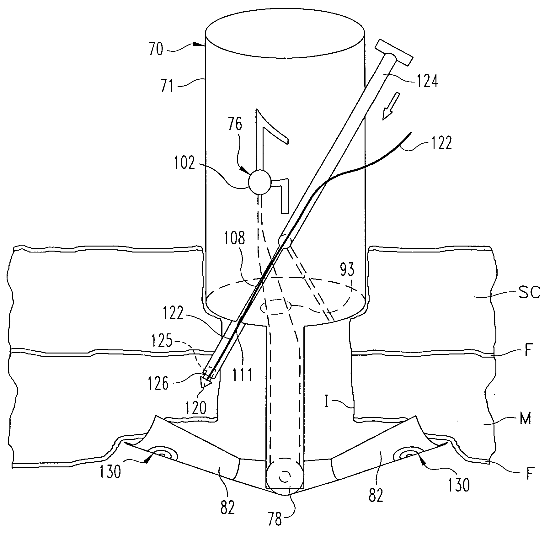
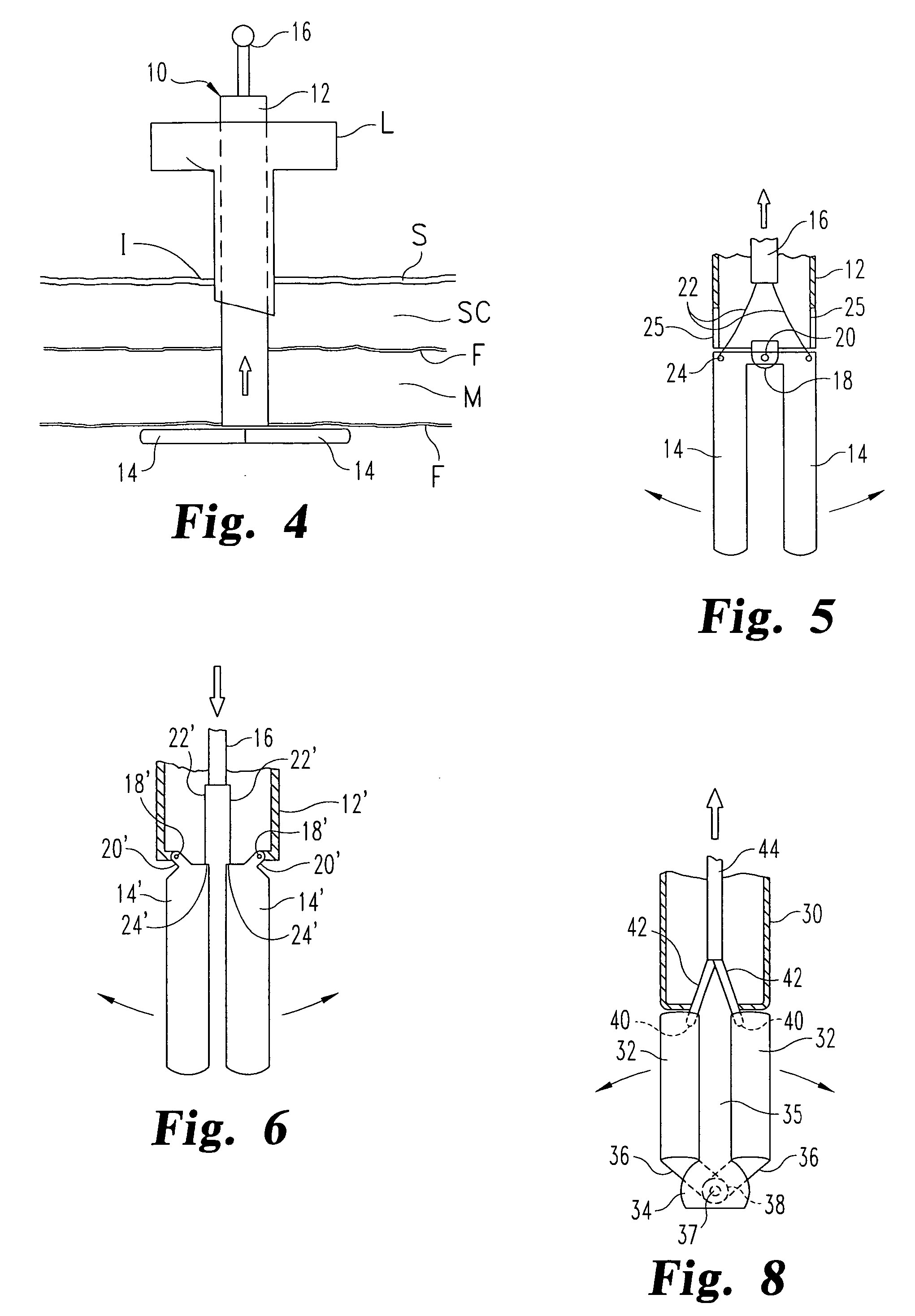
[0013] In the preferred embodiment, the stabilizer elements include a pair of wings pivotably mounted to the distal end of the tubular body. The wings are supported on an axle extending between a pair of legs extending from the lower edge of the tubular body. A torsion spring helps bias the legs to their extended or stabilizing position. An actuator is provided that is accessible outside the patient that can be manipulated to move the stabilizer elements from their insertion position to their stabilizing position, and then to a removal position when it is desired to withdraw the device from the incision.
[0017] In accordance with one embodiment of the invention, a method is provided for assisting in the closure of an incision in body tissue comprising: introducing a tubular body through the incision; deploying a stabilizing element between the body tissue and body structure adjacent the incision; and with the stabilizing element deployed, pulling the tubular body to retract the body tissue from the body structures. With the body tissue retracted, the adjacent body structures can be more easily visualized.
[0018] In another aspect, the method contemplates the stabilizing elements includes forming a space between the stabilizing elements and a lower edge of the tubular body sufficient for ingress of the body tissue into the space. This aspect facilitates advancement of a needle tip carrying a suture therethrough to be captured by the stabilizing element. Thus, a further aspect of the inventive method comprises advancing the needle tip and suture from the tubular body, through body tissue between the tubular body and toward the stabilizing element, and then capturing the needle tip with the suture at a free end of the stabilizing element. Once captured, the stabilizing elements can be moved to a removal position with the needle tip captured at the free end thereof. The tubular body is withdrawn from the incision with the needle tip captured so that the suture forms a ligature through the body tissue at the incision.
[0019] It is one object of the present invention to provide a closure device that facilitates the closure of an incision through body tissue of a patient. Another object of the invention is achieved by features that allow for a minimal number of steps to introduce and operate the closure device to pass sutures through the body tissue.

Problems solved by technology

Although trocars are widely used to puncture the abdominal wall as a first step in minimally invasive surgical techniques, such use creates several clinical problems.
The very small size of the incision and the somewhat awkward access to the interior facies of the tissues surrounding the incision make closure of the incision problematic and time consuming.
If the needle distance from the incision is too small, an insufficient amount of tissue will be secured with a consequent risk of inadequate closure of the surgical defect.
This may result in subsequent herniation of the omentum or bowel.
However, if needle distance from the point of the original trocar incision is too great, incision closure will result in excessive tissue being grasped, and the patient will be left with an unsightly “knot” of tissue.
Aside from attendant awkwardness and the problems resulting therefrom, this method is time-consuming and often produces only marginal closure integrity.
Another difficulty associated with this mode for closure is associated with obese patients who present considerable fat in the abdominal region.
Because the abdominal wall of an obese patient may be several inches thick, it is extremely difficult, tedious and time consuming to approximate the fascial tissues with a suture.
Often times, following removal of a large trocar, the puncture site needs to be enlarged to accomplish this, thus negating some of the advantages of endoscopic surgery previously discussed.
The difficulty with this technique is that the edges of the fascia are not easily visualized, with the result that tying the ligature may or may not effectively re-approximate the edges of the fascia.
Certainly the peritoneal defect is not effectively closed by this approach because the suture is not placed deeply enough.
Failure to make complete closure of the incision entails a significant risk of delayed bleeding (occurring after the abdomen is deflated and the tamponading effect of the inflated abdomen ceases), or the possibility of herniation of either omentum or bowel into the subcutaneous opening.
This approach is rarely used because it is cumbersome, requires a high level of skill, and is still not optimal as it ensures only that the peritoneum is closed, closure of the more exterior fascia being purely speculative.

Method used


Image

  • Laparoscopic port site closure tool
  • Laparoscopic port site closure tool
  • Laparoscopic port site closure tool

Examples


Embodiment Construction

[0041] For the purposes of promoting an understanding of the principles of the invention, reference will now be made to the embodiments illustrated in the drawings and described in the following written specification. It is understood that no limitation to the scope of the invention is thereby intended. It is further understood that the present invention includes any alterations and modifications to the illustrated embodiments and includes further applications of the principles of the invention as would normally occur to one skilled in the art to which this invention pertains.

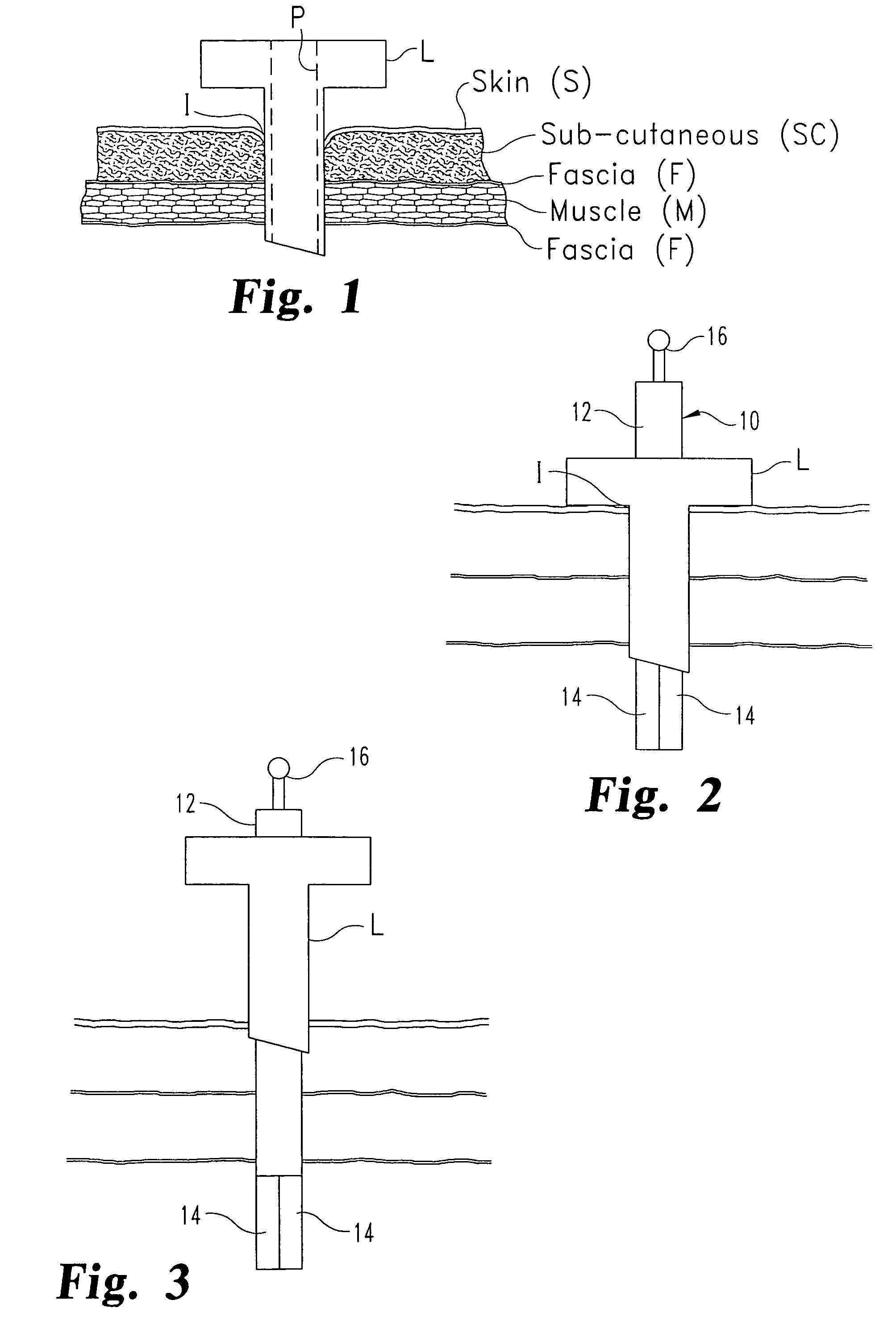
[0042] One phase of a typical laparoscopic procedure is shown in FIG. 1. In particular, a laparoscopic tool L defining a port P is extended into a small incision I. The incision passes through multiple tissue layers for access to the patient's abdomen, for instance. Thus, the laparoscopic tool extends through the skin, subcutaneous and muscle layers, as well as the various fascia layers to provide access to th...


PUM


Abstract

A device is provided to assist in closing an incision through body tissue that comprises a tubular body sized for introduction through the incision and carrying a plurality of stabilizer elements at the distal end thereof. The stabilizer elements are configured to be deployed within the body cavity to engage the lowermost tissue layer. In one method, the device is pulled with the stabilizer elements deployed to retract the body tissue away from adjacent body structure. In another method, a plurality of needle tips carrying sutures are guided through the body tissue to a retention device at the ends of the stabilizer elements. With the needle tips captured in the retention devices, the device is withdrawn from the incision so that the sutures form ligatures that can be tied off to close the incision.

Description

CROSS-REFERENCE TO RELATED APPLICATIONS [0001] This application claims priority to co-pending provisional application No. 60 / 598,798, filed on Aug. 5, 2004, by the present invention. The disclosure and figures of this provisional application are incorporated herein by reference.BACKGROUND OF THE INVENTION [0002] The invention relates generally to surgical suturing devices, and more particularly to intra-abdominal suturing devices designed for closing puncture wounds created by surgical trocars and similar puncturing devices. [0003] Minimally invasive surgery is a revolutionary new technique that has replaced many standard invasive surgical operations requiring large incisions with operations utilizing very small incisions. In this technique, access to the surgical field is made through very small incisions (generally 5-18 mm in diameter) via a surgical trocar. These trocars typically have a diameter of, for example, between 3 mm and 30 mm and a length of about 150 mm (6 inches). Tub...

Claims


Application Information

Patent Timeline
Patent Type & Authority Applications(United States)
IPC IPC(8): A61B17/04
CPCA61B17/0057A61B17/0482A61B17/0483A61B2017/06057A61B2017/00637A61B2017/00663A61B17/06066
Inventor BENNETT, RICHARD M. III
Owner TELEFLEX MEDICAL INC
Learn More