
Method and system for controlled scanning, imaging and/or therapy

a controlled scanning and imaging technology, applied in the field of imaging and treatment systems, can solve the problems of lack of precision and repeatability in the interrogation of 3-d volume, -hand-to-hand scanning, and inability to provide such features in prior art methodologies, and achieve accurate and computationally efficient three-dimensional imaging. , the effect of facilitating three-dimensional imaging

Active Publication Date: 2005-11-17
GUIDED THERAPY SYSTEMS LLC
View PDF99 Cites 185 Cited by
  • Summary
  • Abstract
  • Description
  • Claims
  • Application Information

AI Technical Summary

Benefits of technology

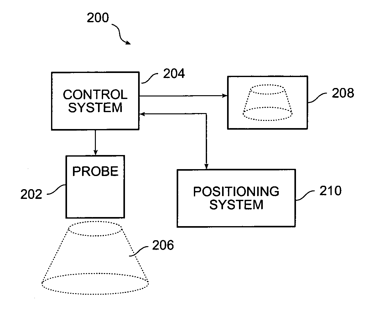
[0006] A method and system for controlled scanning, imaging and / or therapy are provided. In accordance with one aspect, an exemplary method and system are configured to suitably control an imaging probe within a one-degree of freedom. With such control, an exemplary method and system can facilitate three-dimensional imaging. For example, an exemplary method and system can enable multiple two-dimensional image planes to be collected in a manner to provide an accurate and computationally efficient three-dimensional image reconstruction while providing the user with a user-friendly mechanism for acquiring three-dimensional images. In accordance with another aspect of the present invention, an exemplary method and system can allow therapeutic treatment to occur along a prescribed path or pattern. For example, treatments that would normally occur at a single point in space become a line or other guided path after scanning in the controlled pattern, while line treatments scanned along a path can suitably become a matrix of treatments.

Problems solved by technology

Unfortunately, prior art methodologies cannot provide such features.
For example, one shortcoming of so-called “free-hand” 3-D scanning is the lack of precision and repeatability in which the 3-D volume is interrogated due to spatially and temporally imprecise angular and linear displacements.
As a result a number of pitfalls exist.
Second, even if the volume is adequately sampled, the random nature of the input data orientation requires excessive mathematical interpolations to compute a 3-D image in a uniform output grid.
Third, if no sensors are used image frame correlation methods cannot accurately ascertain the relative orientation of image planes.
Finally, even if six-degree-of-freedom sensors are utilized, such sensors are expensive and have limited range.
However, such mechanisms can be extremely cumbersome and unwieldy for human scanning and may pose safety hazards if designed improperly.

Method used


Image

  • Method and system for controlled scanning, imaging and/or therapy
  • Method and system for controlled scanning, imaging and/or therapy
  • Method and system for controlled scanning, imaging and/or therapy

Examples


Embodiment Construction

[0022] The present invention may be described herein in terms of various functional components and processing steps. It should be appreciated that such components and steps may be realized by any number of hardware components configured to perform the specified functions. For example, the present invention may employ various medical treatment devices, visual imaging and display devices, input terminals and the like, which may carry out a variety of functions under the control of one or more control systems or other control devices. In addition, the present invention may be practiced in any number of medical contexts and that the exemplary embodiments relating to an imaging, therapy and / or scanning system as described herein are merely indicative of exemplary applications for the invention. For example, the principles, features and methods discussed may be applied to any medical application. Further, various aspects of the present invention may be suitably applied to other industrial...


PUM


Abstract

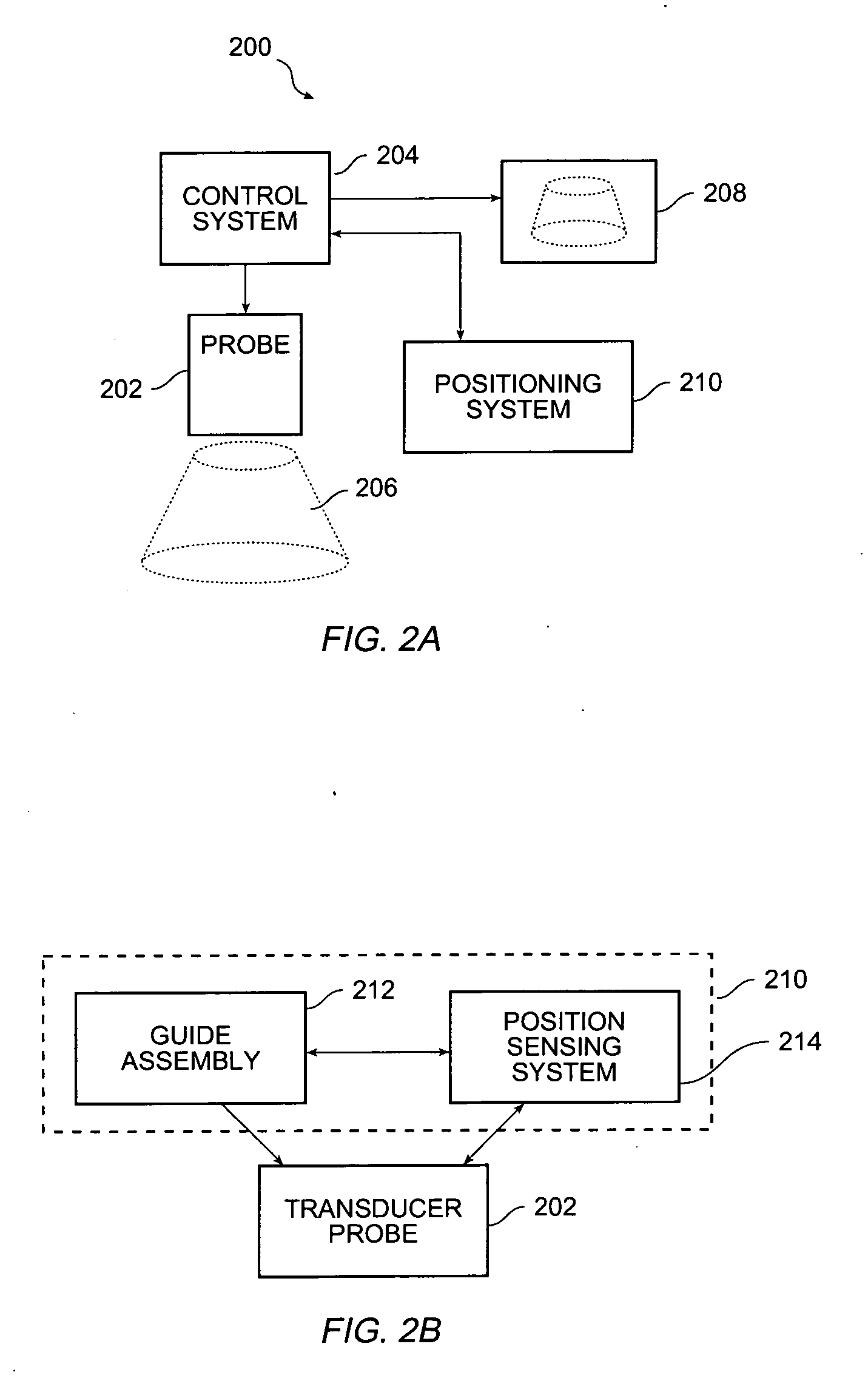
A method and system for three dimensional scanning, imaging and / or therapy are provided. In accordance with one aspect, an exemplary method and system are configured to facilitate controlled scanning within one-degree of freedom. For example, an exemplary method and system can enable multiple two-dimensional image planes to be collected in a manner to provide an accurate and computationally efficient three-dimensional image reconstruction while providing the user with a user-friendly mechanism for acquiring three-dimensional images. In accordance with an exemplary embodiment, an exemplary scanning and imaging system comprises an imaging probe, a control system, a positioning system and a display system. In accordance with an exemplary embodiment, the positioning system comprises a guide assembly and a position sensing system. The guide assembly is configured to provide pure rectilinear or rotational motion of the probe during scanning operation while the position sensing system is configured to detect the direction and position of the probe during scanning.

Description

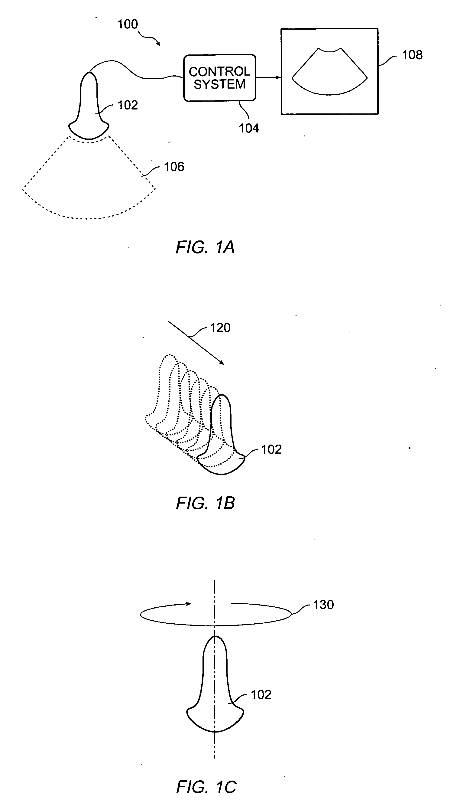
CROSS-REFERENCE TO RELATED APPLICATIONS [0001] This application claim priority from U.S. Provisional Application No. 60 / 570,145, entitled “Method and System for Three-Dimensional Scanning and Imaging” and filed May 12, 2004.FIELD OF INVENTION [0002] The present invention relates to imaging and treatment systems, and in particular to a method and system for controlled scanning, imaging and / or therapy. BACKGROUND OF THE INVENTION [0003] Ultrasound images are typically generated and displayed as two-dimensional (2-D) image slices. For example, with reference to FIG. 1A, a conventional ultrasound imaging system 100 comprising a transducer 102 and a control system 104 are configured to obtain two-dimensional imaging information 106 and display two-dimensional imaging slices 108. However, it is often desirable to acquire a whole volume of data in the form of multiple image planes and render it in a three-dimensional (3-D) format, such as for viewing a fetus. Acquiring multiple image slice...

Claims


Application Information

Patent Timeline
Patent Type & Authority Applications(United States)
IPC IPC(8): A61B8/02A61B8/12A61B8/14
CPCA61B8/14A61B8/483A61B8/4209
Inventor BARTHE, PETER G.SLAYTON, MICHAEL H.
Owner GUIDED THERAPY SYSTEMS LLC
Learn More