
Diagnosis aid apparatus

a technology of diagnostic aid and equipment, applied in the field of diagnostic aid equipment, can solve the problems of difficulty in judgment of algorithm and setup for improving the diagnostic accuracy and the detection rate of cancer and the like, and the accuracy of detection executed by automatic detection processing is only obtained, so as to facilitate adjustment and improve the diagnostic efficiency

Inactive Publication Date: 2005-02-24
KONICA MINOLTA MEDICAL & GRAPHICS INC
View PDF6 Cites 25 Cited by
  • Summary
  • Abstract
  • Description
  • Claims
  • Application Information

AI Technical Summary

Benefits of technology

"The present invention provides a diagnosis aid apparatus for detecting abnormal shadows in medical images. The apparatus includes a display section for showing the medical image, an abnormal shadow candidate detecting section, a detection performance inspecting section, and a data outputting section. The apparatus can adjust the detection performance of the abnormal shadow candidate based on the doctor's skill by displaying the detection performance of the abnormal shadow candidate and the accompanying information of the medical image. This helps to improve the interpretation of the medical image and improve the diagnosis efficiency of the abnormal shadow candidate."

Problems solved by technology

However, in the above-described abnormal shadow processing system, by relating the results of detection processing about a plurality of image information to corrected result after correcting the result, storing and testing them by comparison, an accuracy of detection executed by automatic detection processing is only obtained by statistics processing.
However, these operation are troublesome, and judgment of the algorithm and setup for improving the diagnostic accuracy and the detection rate of cancer and the like, as a whole, not individual, are difficult.

Method used


Image

  • Diagnosis aid apparatus
  • Diagnosis aid apparatus
  • Diagnosis aid apparatus

Examples


Embodiment Construction

[0051] Hereinafter, embodiments of the invention will be explained in detail with reference to FIG. 1 to FIG. 8C, and thus scope of the invention is not limited to the example of illustration.

[0052] First, configuration of the embodiments will be explained.

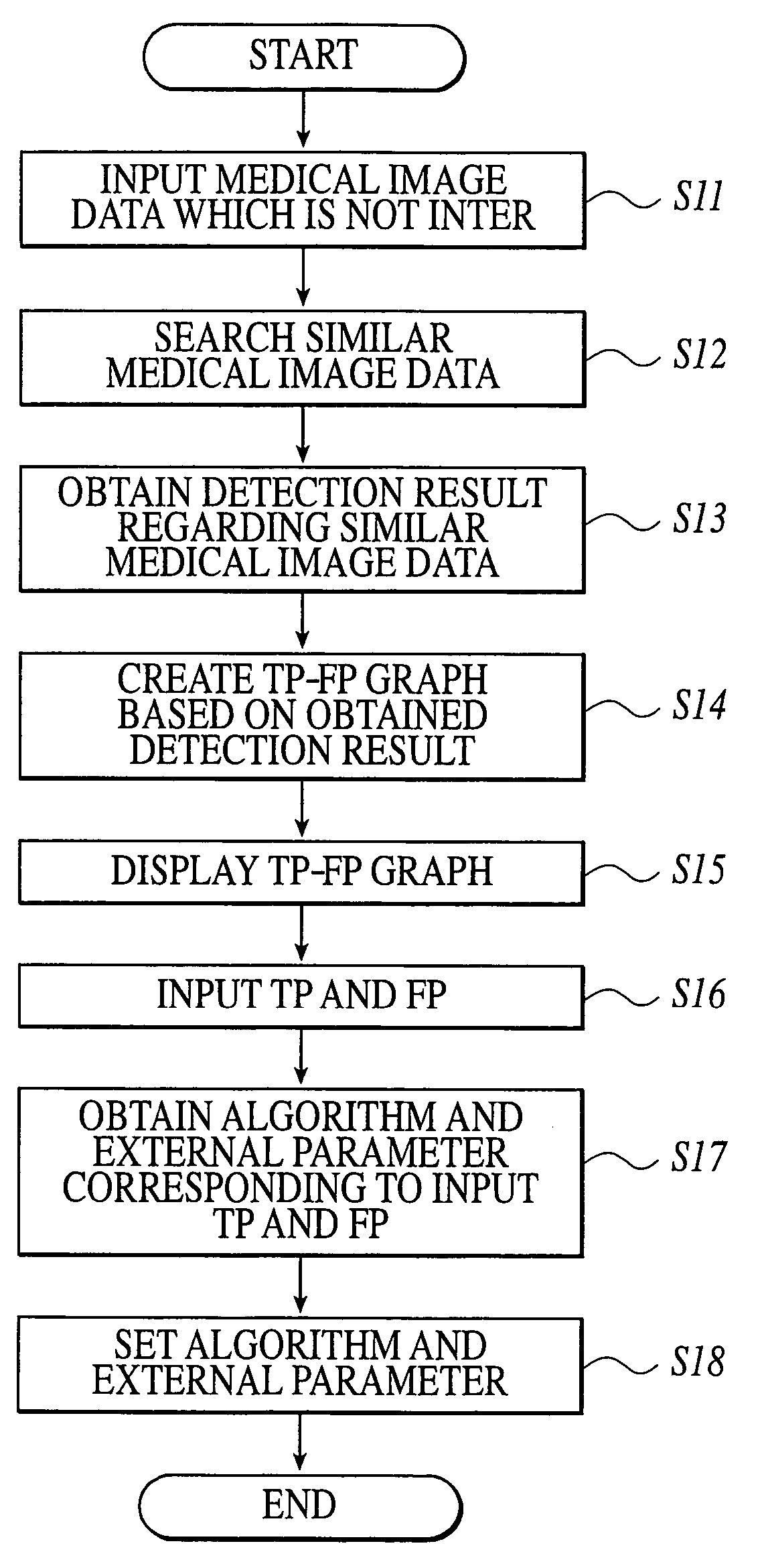
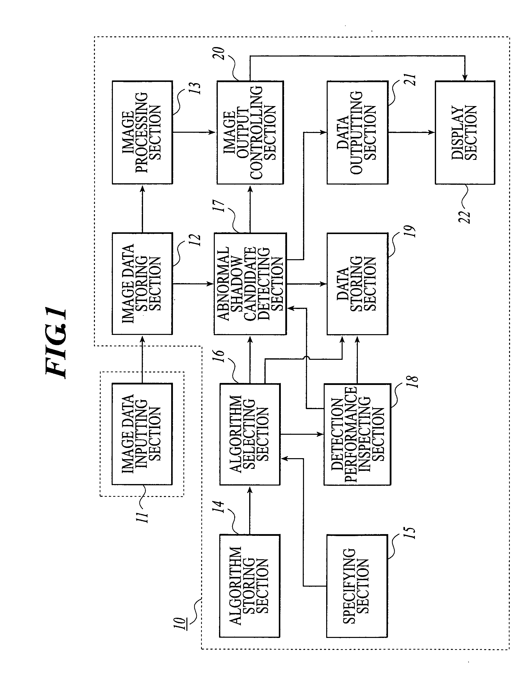
[0053]FIG. 1 is a view showing functional configuration of the diagnosis aid apparatus 10 in the present embodiments. As shown in FIG. 1, the diagnosis aid apparatus 10 connected to a image data inputting section 11 comprises an image data storing section 12, an image processing section 13, an algorithm storing section 14, a specifying section 15, an algorithm selecting section 16, an abnormal shadow candidate detecting section 17, a detection performance inspecting section 18, a data storing section 19, an image output controlling section 20, a data outputting section 21, a display section 22 and the like. Further, in the present embodiments, the diagnosis aid apparatus 10 and the image data inputting section will be explained ...


PUM


Abstract

A diagnosis aid apparatus includes: a display section for displaying a medical image of a photographed subject; an abnormal shadow candidate detecting section for detecting an abnormal shadow candidate from the medical image based on a detection condition; a detection performance inspecting section for inspecting a detection performance in each detection condition of the abnormal shadow candidate by comparing a detection result of the abnormal shadow candidate detected by the abnormal shadow candidate detecting section with accompanying information of the medical image; and a data outputting section for displaying the detection performance of the abnormal shadow candidate, which is inspected by the detection performance inspecting section, on the display section.

Description

BACKGROUND OF THE INVENTION [0001] 1. Field of the Invention [0002] The invention relates to diagnosis aid apparatus which detects the abnormal shadow candidate by making image analysis of a medical image. [0003] 2. Description of Related Art [0004] In a medical field, digitalization of a medical image which is generated by photographing a subject is realized. At a diagnosis, a medical image data generated by CR (Computed Radiography), CT (Computed Tomography), MRI (Magnetic Resonance Imaging), US (Ultrasound System) and the like, is displayed on a monitor and subsequently, a doctor interprets the medical image displayed on the monitor and observes condition and change of lesion. [0005] According to an earlier development, for the purpose of reducing load on a doctor, medical image processing apparatus called Computed-Aided Diagnosis (hereinafter, referred to as a CAD) which detects lesion as the abnormal shadow candidate automatically is developed. This detection processing of the ...

Claims


Application Information

Patent Timeline
Patent Type & Authority Applications(United States)
IPC IPC(8): G01R33/32A61B5/055A61B6/00A61B6/03A61B8/00G06K9/00G06T1/00G06T7/00
CPCG06T7/0012
Inventor YAMANAKA, KENJI
Owner KONICA MINOLTA MEDICAL & GRAPHICS INC
Learn More